Iron »
PDB 5why-5xbp »
5ww8 »
Iron in PDB 5ww8: Crystal Structure of the Second Dna-Binding Protein Under Starvation From Mycobacterium Smegmatis Soaked with Iron in the Ratio of 480 Iron Atoms Per Dodecamer
Protein crystallography data
The structure of Crystal Structure of the Second Dna-Binding Protein Under Starvation From Mycobacterium Smegmatis Soaked with Iron in the Ratio of 480 Iron Atoms Per Dodecamer, PDB code: 5ww8
was solved by
S.M.Williams,
D.Chatterji,
with X-Ray Crystallography technique. A brief refinement statistics is given in the table below:
| Resolution Low / High (Å) | 30.00 / 2.04 |
| Space group | H 3 2 |
| Cell size a, b, c (Å), α, β, γ (°) | 89.700, 89.700, 420.842, 90.00, 90.00, 120.00 |
| R / Rfree (%) | 17.7 / 21 |
Other elements in 5ww8:
The structure of Crystal Structure of the Second Dna-Binding Protein Under Starvation From Mycobacterium Smegmatis Soaked with Iron in the Ratio of 480 Iron Atoms Per Dodecamer also contains other interesting chemical elements:
| Magnesium | (Mg) | 2 atoms |
| Chlorine | (Cl) | 2 atoms |
Iron Binding Sites:
The binding sites of Iron atom in the Crystal Structure of the Second Dna-Binding Protein Under Starvation From Mycobacterium Smegmatis Soaked with Iron in the Ratio of 480 Iron Atoms Per Dodecamer
(pdb code 5ww8). This binding sites where shown within
5.0 Angstroms radius around Iron atom.
In total 8 binding sites of Iron where determined in the Crystal Structure of the Second Dna-Binding Protein Under Starvation From Mycobacterium Smegmatis Soaked with Iron in the Ratio of 480 Iron Atoms Per Dodecamer, PDB code: 5ww8:
Jump to Iron binding site number: 1; 2; 3; 4; 5; 6; 7; 8;
In total 8 binding sites of Iron where determined in the Crystal Structure of the Second Dna-Binding Protein Under Starvation From Mycobacterium Smegmatis Soaked with Iron in the Ratio of 480 Iron Atoms Per Dodecamer, PDB code: 5ww8:
Jump to Iron binding site number: 1; 2; 3; 4; 5; 6; 7; 8;
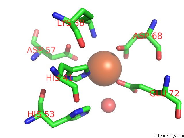
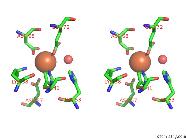
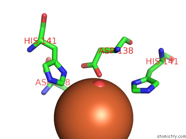
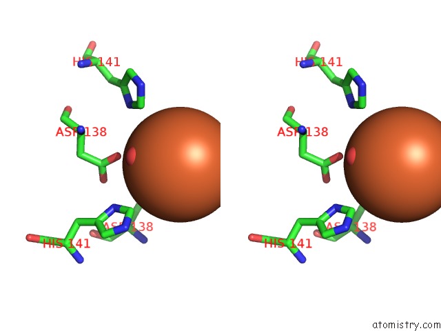
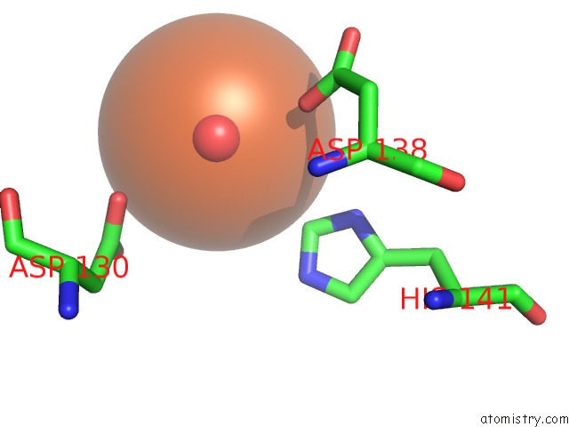
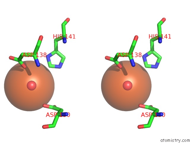
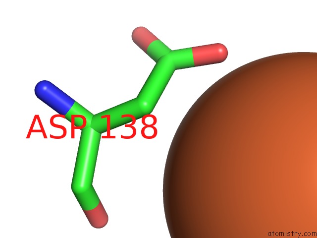
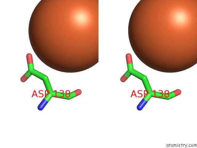
Iron binding site 1 out of 8 in 5ww8
Go back to
Iron binding site 1 out
of 8 in the Crystal Structure of the Second Dna-Binding Protein Under Starvation From Mycobacterium Smegmatis Soaked with Iron in the Ratio of 480 Iron Atoms Per Dodecamer
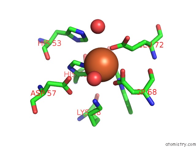
Mono view
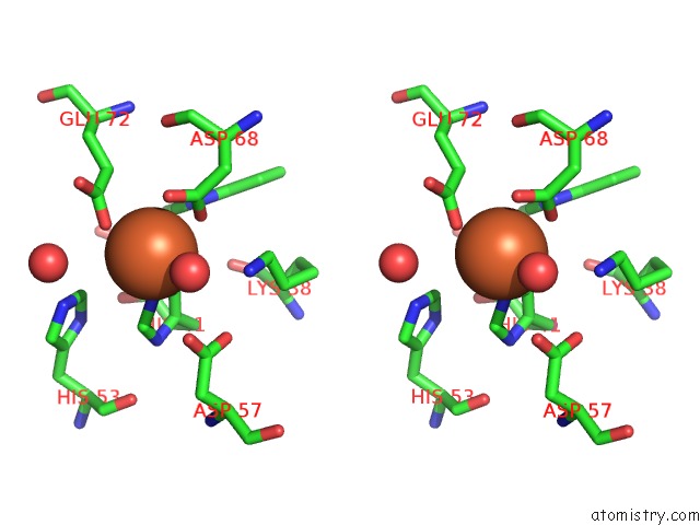
Stereo pair view
Mono view
Stereo pair view
A full contact list of Iron with other atoms in the Fe binding
site number 1 of Crystal Structure of the Second Dna-Binding Protein Under Starvation From Mycobacterium Smegmatis Soaked with Iron in the Ratio of 480 Iron Atoms Per Dodecamer within 5.0Å range:
|
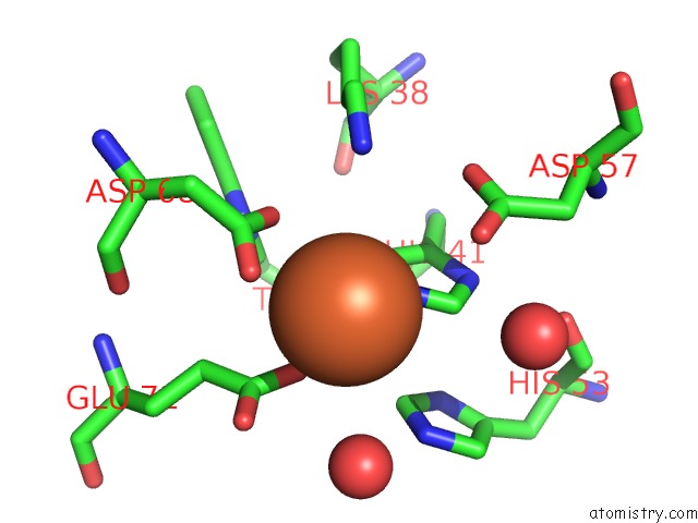
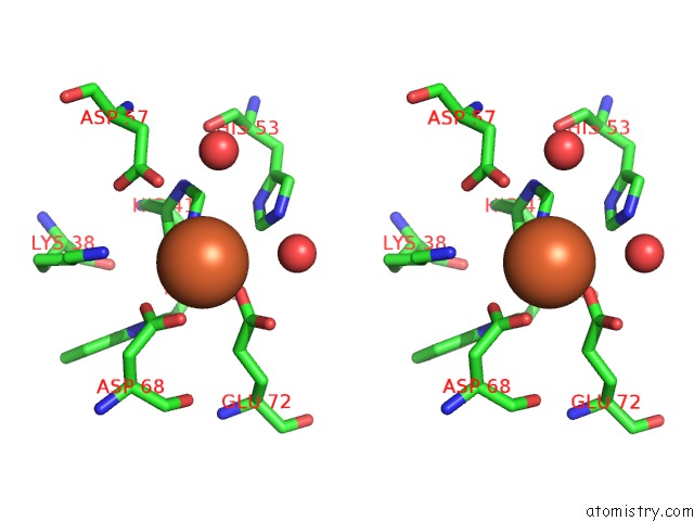
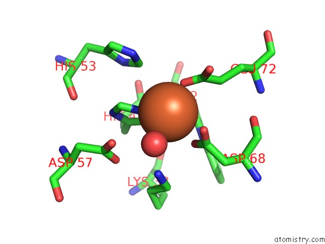
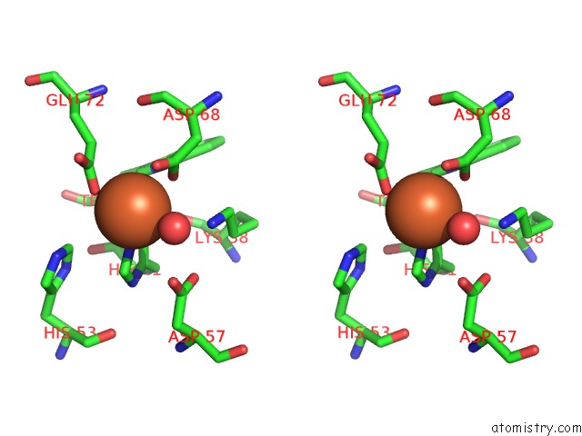
Iron binding site 2 out of 8 in 5ww8
Go back to
Iron binding site 2 out
of 8 in the Crystal Structure of the Second Dna-Binding Protein Under Starvation From Mycobacterium Smegmatis Soaked with Iron in the Ratio of 480 Iron Atoms Per Dodecamer
Mono view
Stereo pair view
Mono view
Stereo pair view
A full contact list of Iron with other atoms in the Fe binding
site number 2 of Crystal Structure of the Second Dna-Binding Protein Under Starvation From Mycobacterium Smegmatis Soaked with Iron in the Ratio of 480 Iron Atoms Per Dodecamer within 5.0Å range:
|
Iron binding site 3 out of 8 in 5ww8
Go back to
Iron binding site 3 out
of 8 in the Crystal Structure of the Second Dna-Binding Protein Under Starvation From Mycobacterium Smegmatis Soaked with Iron in the Ratio of 480 Iron Atoms Per Dodecamer
Mono view
Stereo pair view
Mono view
Stereo pair view
A full contact list of Iron with other atoms in the Fe binding
site number 3 of Crystal Structure of the Second Dna-Binding Protein Under Starvation From Mycobacterium Smegmatis Soaked with Iron in the Ratio of 480 Iron Atoms Per Dodecamer within 5.0Å range:
|
Iron binding site 4 out of 8 in 5ww8
Go back to
Iron binding site 4 out
of 8 in the Crystal Structure of the Second Dna-Binding Protein Under Starvation From Mycobacterium Smegmatis Soaked with Iron in the Ratio of 480 Iron Atoms Per Dodecamer
Mono view
Stereo pair view
Mono view
Stereo pair view
A full contact list of Iron with other atoms in the Fe binding
site number 4 of Crystal Structure of the Second Dna-Binding Protein Under Starvation From Mycobacterium Smegmatis Soaked with Iron in the Ratio of 480 Iron Atoms Per Dodecamer within 5.0Å range:
|
Iron binding site 5 out of 8 in 5ww8
Go back to
Iron binding site 5 out
of 8 in the Crystal Structure of the Second Dna-Binding Protein Under Starvation From Mycobacterium Smegmatis Soaked with Iron in the Ratio of 480 Iron Atoms Per Dodecamer
Mono view
Stereo pair view
Mono view
Stereo pair view
A full contact list of Iron with other atoms in the Fe binding
site number 5 of Crystal Structure of the Second Dna-Binding Protein Under Starvation From Mycobacterium Smegmatis Soaked with Iron in the Ratio of 480 Iron Atoms Per Dodecamer within 5.0Å range:
|
Iron binding site 6 out of 8 in 5ww8
Go back to
Iron binding site 6 out
of 8 in the Crystal Structure of the Second Dna-Binding Protein Under Starvation From Mycobacterium Smegmatis Soaked with Iron in the Ratio of 480 Iron Atoms Per Dodecamer
Mono view
Stereo pair view
Mono view
Stereo pair view
A full contact list of Iron with other atoms in the Fe binding
site number 6 of Crystal Structure of the Second Dna-Binding Protein Under Starvation From Mycobacterium Smegmatis Soaked with Iron in the Ratio of 480 Iron Atoms Per Dodecamer within 5.0Å range:
|
Iron binding site 7 out of 8 in 5ww8
Go back to
Iron binding site 7 out
of 8 in the Crystal Structure of the Second Dna-Binding Protein Under Starvation From Mycobacterium Smegmatis Soaked with Iron in the Ratio of 480 Iron Atoms Per Dodecamer
Mono view
Stereo pair view
Mono view
Stereo pair view
A full contact list of Iron with other atoms in the Fe binding
site number 7 of Crystal Structure of the Second Dna-Binding Protein Under Starvation From Mycobacterium Smegmatis Soaked with Iron in the Ratio of 480 Iron Atoms Per Dodecamer within 5.0Å range:
|
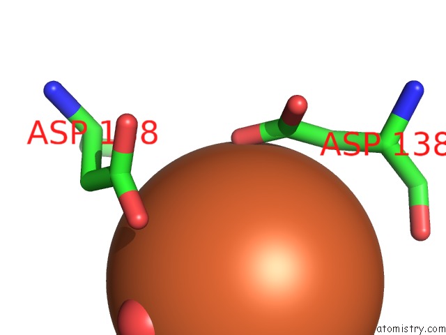
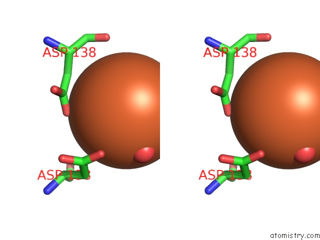
Iron binding site 8 out of 8 in 5ww8
Go back to
Iron binding site 8 out
of 8 in the Crystal Structure of the Second Dna-Binding Protein Under Starvation From Mycobacterium Smegmatis Soaked with Iron in the Ratio of 480 Iron Atoms Per Dodecamer
Mono view
Stereo pair view
Mono view
Stereo pair view
A full contact list of Iron with other atoms in the Fe binding
site number 8 of Crystal Structure of the Second Dna-Binding Protein Under Starvation From Mycobacterium Smegmatis Soaked with Iron in the Ratio of 480 Iron Atoms Per Dodecamer within 5.0Å range:
|
Reference:
S.M.Williams,
D.Chatterji.
Flexible Aspartates Propel Iron to the Ferroxidation Sites Along Pathways Stabilized By A Conserved Arginine in Dps Proteins From Mycobacterium Smegmatis Metallomics V. 9 685 2017.
ISSN: ESSN 1756-591X
PubMed: 28418062
DOI: 10.1039/C7MT00008A
Page generated: Tue Aug 6 11:23:06 2024
ISSN: ESSN 1756-591X
PubMed: 28418062
DOI: 10.1039/C7MT00008A
Last articles
Fe in 2YXOFe in 2YRS
Fe in 2YXC
Fe in 2YNM
Fe in 2YVJ
Fe in 2YP1
Fe in 2YU2
Fe in 2YU1
Fe in 2YQB
Fe in 2YOO