Iron »
PDB 5xzi-5ylw »
5y1i »
Iron in PDB 5y1i: The Crystal Structure of Gfsf
Protein crystallography data
The structure of The Crystal Structure of Gfsf, PDB code: 5y1i
was solved by
A.Miyanaga,
F.Kudo,
T.Eguchi,
with X-Ray Crystallography technique. A brief refinement statistics is given in the table below:
| Resolution Low / High (Å) | 50.49 / 2.00 |
| Space group | P 21 21 21 |
| Cell size a, b, c (Å), α, β, γ (°) | 67.080, 102.870, 114.430, 90.00, 90.00, 90.00 |
| R / Rfree (%) | 19.4 / 23.6 |
Iron Binding Sites:
The binding sites of Iron atom in the The Crystal Structure of Gfsf
(pdb code 5y1i). This binding sites where shown within
5.0 Angstroms radius around Iron atom.
In total 2 binding sites of Iron where determined in the The Crystal Structure of Gfsf, PDB code: 5y1i:
Jump to Iron binding site number: 1; 2;
In total 2 binding sites of Iron where determined in the The Crystal Structure of Gfsf, PDB code: 5y1i:
Jump to Iron binding site number: 1; 2;
Iron binding site 1 out of 2 in 5y1i
Go back to
Iron binding site 1 out
of 2 in the The Crystal Structure of Gfsf

Mono view
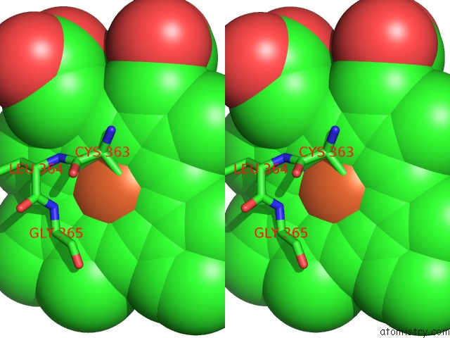
Stereo pair view
Mono view
Stereo pair view
A full contact list of Iron with other atoms in the Fe binding
site number 1 of The Crystal Structure of Gfsf within 5.0Å range:
|
Iron binding site 2 out of 2 in 5y1i
Go back to
Iron binding site 2 out
of 2 in the The Crystal Structure of Gfsf
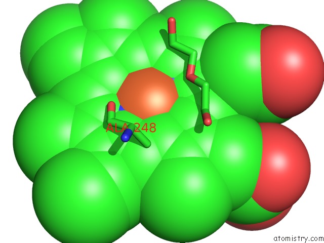
Mono view

Stereo pair view
Mono view
Stereo pair view
A full contact list of Iron with other atoms in the Fe binding
site number 2 of The Crystal Structure of Gfsf within 5.0Å range:
|
Reference:
A.Miyanaga,
R.Takayanagi,
T.Furuya,
A.Kawamata,
T.Itagaki,
Y.Iwabuchi,
N.Kanoh,
F.Kudo,
T.Eguchi.
Substrate Recognition By A Dual-Function P450 Monooxygenase Gfsf Involved in Fd-891 Biosynthesis Chembiochem V. 18 2179 2017.
ISSN: ESSN 1439-7633
PubMed: 28869713
DOI: 10.1002/CBIC.201700429
Page generated: Tue Aug 6 12:36:34 2024
ISSN: ESSN 1439-7633
PubMed: 28869713
DOI: 10.1002/CBIC.201700429
Last articles
Zn in 9J0NZn in 9J0O
Zn in 9J0P
Zn in 9FJX
Zn in 9EKB
Zn in 9C0F
Zn in 9CAH
Zn in 9CH0
Zn in 9CH3
Zn in 9CH1