Iron »
PDB 5xzi-5ylw »
5y5g »
Iron in PDB 5y5g: Structure of Cytochrome P450NOR in No-Bound State: Damaged By Low-Dose (0.72 Mgy) X-Ray
Enzymatic activity of Structure of Cytochrome P450NOR in No-Bound State: Damaged By Low-Dose (0.72 Mgy) X-Ray
All present enzymatic activity of Structure of Cytochrome P450NOR in No-Bound State: Damaged By Low-Dose (0.72 Mgy) X-Ray:
1.7.1.14;
1.7.1.14;
Protein crystallography data
The structure of Structure of Cytochrome P450NOR in No-Bound State: Damaged By Low-Dose (0.72 Mgy) X-Ray, PDB code: 5y5g
was solved by
T.Tosha,
T.Nomura,
T.Nishida,
G.Ueno,
H.Murakami,
K.Yamashita,
K.Hirata,
M.Yamamoto,
H.Ago,
H.Sugimoto,
Y.Shiro,
M.Kubo,
with X-Ray Crystallography technique. A brief refinement statistics is given in the table below:
| Resolution Low / High (Å) | 26.97 / 1.36 |
| Space group | P 21 21 21 |
| Cell size a, b, c (Å), α, β, γ (°) | 55.189, 75.477, 101.668, 90.00, 90.00, 90.00 |
| R / Rfree (%) | 13.3 / 16.6 |
Iron Binding Sites:
The binding sites of Iron atom in the Structure of Cytochrome P450NOR in No-Bound State: Damaged By Low-Dose (0.72 Mgy) X-Ray
(pdb code 5y5g). This binding sites where shown within
5.0 Angstroms radius around Iron atom.
In total only one binding site of Iron was determined in the Structure of Cytochrome P450NOR in No-Bound State: Damaged By Low-Dose (0.72 Mgy) X-Ray, PDB code: 5y5g:
In total only one binding site of Iron was determined in the Structure of Cytochrome P450NOR in No-Bound State: Damaged By Low-Dose (0.72 Mgy) X-Ray, PDB code: 5y5g:
Iron binding site 1 out of 1 in 5y5g
Go back to
Iron binding site 1 out
of 1 in the Structure of Cytochrome P450NOR in No-Bound State: Damaged By Low-Dose (0.72 Mgy) X-Ray
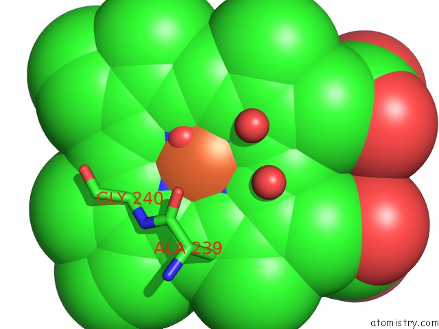
Mono view
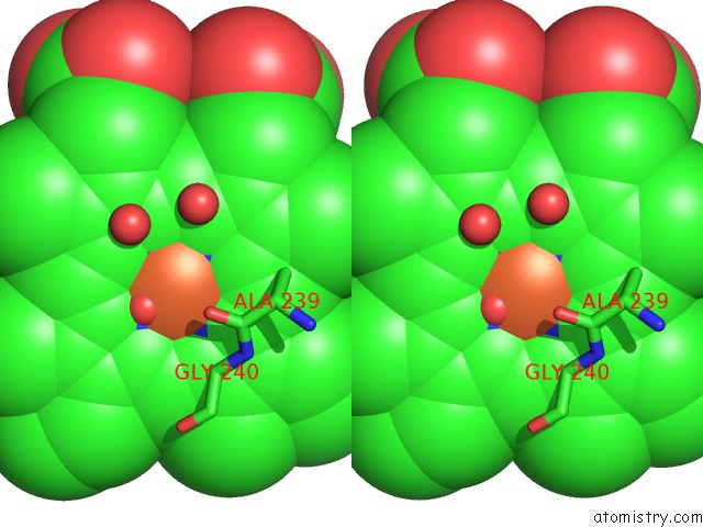
Stereo pair view
Mono view
Stereo pair view
A full contact list of Iron with other atoms in the Fe binding
site number 1 of Structure of Cytochrome P450NOR in No-Bound State: Damaged By Low-Dose (0.72 Mgy) X-Ray within 5.0Å range:
|
Reference:
T.Tosha,
T.Nomura,
T.Nishida,
N.Saeki,
K.Okubayashi,
R.Yamagiwa,
M.Sugahara,
T.Nakane,
K.Yamashita,
K.Hirata,
G.Ueno,
T.Kimura,
T.Hisano,
K.Muramoto,
H.Sawai,
H.Takeda,
E.Mizohata,
A.Yamashita,
Y.Kanematsu,
Y.Takano,
E.Nango,
R.Tanaka,
O.Nureki,
O.Shoji,
Y.Ikemoto,
H.Murakami,
S.Owada,
K.Tono,
M.Yabashi,
M.Yamamoto,
H.Ago,
S.Iwata,
H.Sugimoto,
Y.Shiro,
M.Kubo.
Capturing An Initial Intermediate During the P450NOR Enzymatic Reaction Using Time-Resolved Xfel Crystallography and Caged-Substrate. Nat Commun V. 8 1585 2017.
ISSN: ESSN 2041-1723
PubMed: 29147002
DOI: 10.1038/S41467-017-01702-1
Page generated: Tue Aug 6 12:38:14 2024
ISSN: ESSN 2041-1723
PubMed: 29147002
DOI: 10.1038/S41467-017-01702-1
Last articles
Zn in 9J0NZn in 9J0O
Zn in 9J0P
Zn in 9FJX
Zn in 9EKB
Zn in 9C0F
Zn in 9CAH
Zn in 9CH0
Zn in 9CH3
Zn in 9CH1