Iron »
PDB 5xzi-5ylw »
5y6k »
Iron in PDB 5y6k: Human Serum Trnasferrin Bound to A Fluorescent Probe
Protein crystallography data
The structure of Human Serum Trnasferrin Bound to A Fluorescent Probe, PDB code: 5y6k
was solved by
N.Jiang,
T.Cheng,
M.Wang,
G.C.F.Chan,
L.Jin,
H.Li,
H.Sun,
with X-Ray Crystallography technique. A brief refinement statistics is given in the table below:
| Resolution Low / High (Å) | 103.29 / 2.86 |
| Space group | C 2 2 21 |
| Cell size a, b, c (Å), α, β, γ (°) | 138.012, 155.752, 107.554, 90.00, 90.00, 90.00 |
| R / Rfree (%) | 15.4 / 23.5 |
Iron Binding Sites:
The binding sites of Iron atom in the Human Serum Trnasferrin Bound to A Fluorescent Probe
(pdb code 5y6k). This binding sites where shown within
5.0 Angstroms radius around Iron atom.
In total 2 binding sites of Iron where determined in the Human Serum Trnasferrin Bound to A Fluorescent Probe, PDB code: 5y6k:
Jump to Iron binding site number: 1; 2;
In total 2 binding sites of Iron where determined in the Human Serum Trnasferrin Bound to A Fluorescent Probe, PDB code: 5y6k:
Jump to Iron binding site number: 1; 2;
Iron binding site 1 out of 2 in 5y6k
Go back to
Iron binding site 1 out
of 2 in the Human Serum Trnasferrin Bound to A Fluorescent Probe
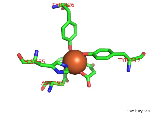
Mono view
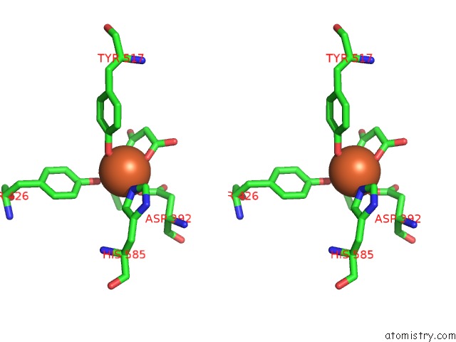
Stereo pair view
Mono view
Stereo pair view
A full contact list of Iron with other atoms in the Fe binding
site number 1 of Human Serum Trnasferrin Bound to A Fluorescent Probe within 5.0Å range:
|
Iron binding site 2 out of 2 in 5y6k
Go back to
Iron binding site 2 out
of 2 in the Human Serum Trnasferrin Bound to A Fluorescent Probe
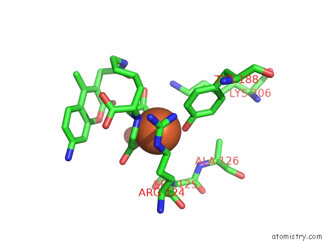
Mono view
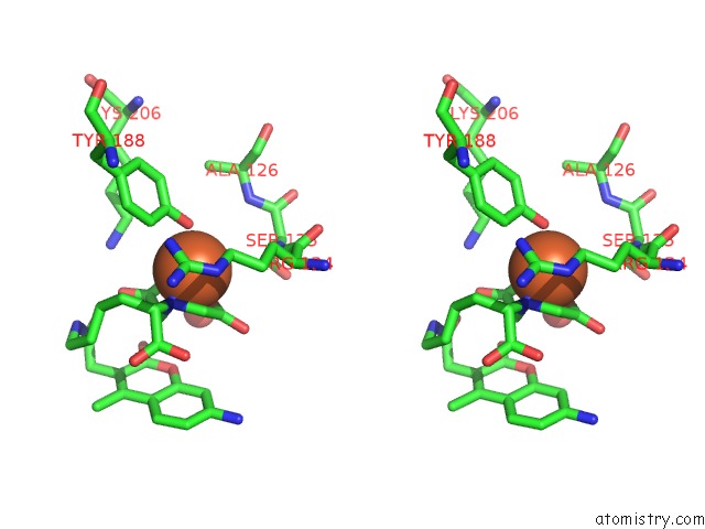
Stereo pair view
Mono view
Stereo pair view
A full contact list of Iron with other atoms in the Fe binding
site number 2 of Human Serum Trnasferrin Bound to A Fluorescent Probe within 5.0Å range:
|
Reference:
N.Jiang,
T.Cheng,
M.Wang,
G.C.Chan,
L.Jin,
H.Li,
H.Sun.
Tracking Iron-Associated Proteomes in Pathogens By A Fluorescence Approach. Metallomics V. 10 77 2018.
ISSN: ESSN 1756-591X
PubMed: 29323384
DOI: 10.1039/C7MT00275K
Page generated: Wed Aug 6 03:20:52 2025
ISSN: ESSN 1756-591X
PubMed: 29323384
DOI: 10.1039/C7MT00275K
Last articles
Fe in 7AQSFe in 7AQR
Fe in 7AIZ
Fe in 7AK6
Fe in 7AK5
Fe in 7AQQ
Fe in 7AO7
Fe in 7ANV
Fe in 7ANT
Fe in 7AKT