Iron »
PDB 5zm9-6air »
5zze »
Iron in PDB 5zze: Crystal Structure of Horse Myoglobin Crystallized By Ammonium Sulfate
Protein crystallography data
The structure of Crystal Structure of Horse Myoglobin Crystallized By Ammonium Sulfate, PDB code: 5zze
was solved by
M.Kitahara,
S.Fudo,
T.Yoneda,
M.Nukaga,
T.Hoshino,
with X-Ray Crystallography technique. A brief refinement statistics is given in the table below:
| Resolution Low / High (Å) | 33.76 / 1.42 |
| Space group | P 1 21 1 |
| Cell size a, b, c (Å), α, β, γ (°) | 35.264, 28.588, 62.822, 90.00, 105.75, 90.00 |
| R / Rfree (%) | 20.2 / 24.8 |
Iron Binding Sites:
The binding sites of Iron atom in the Crystal Structure of Horse Myoglobin Crystallized By Ammonium Sulfate
(pdb code 5zze). This binding sites where shown within
5.0 Angstroms radius around Iron atom.
In total only one binding site of Iron was determined in the Crystal Structure of Horse Myoglobin Crystallized By Ammonium Sulfate, PDB code: 5zze:
In total only one binding site of Iron was determined in the Crystal Structure of Horse Myoglobin Crystallized By Ammonium Sulfate, PDB code: 5zze:
Iron binding site 1 out of 1 in 5zze
Go back to
Iron binding site 1 out
of 1 in the Crystal Structure of Horse Myoglobin Crystallized By Ammonium Sulfate
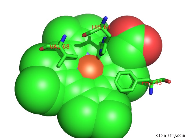
Mono view
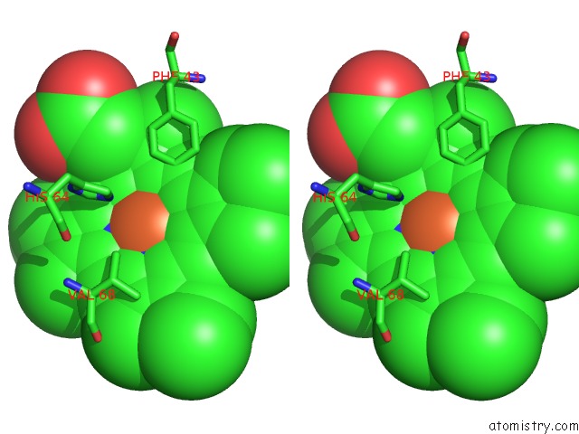
Stereo pair view
Mono view
Stereo pair view
A full contact list of Iron with other atoms in the Fe binding
site number 1 of Crystal Structure of Horse Myoglobin Crystallized By Ammonium Sulfate within 5.0Å range:
|
Reference:
M.Kitahara,
S.Fudo,
T.Yoneda,
M.Nukaga,
T.Hoshino.
Anisotropic Distribution of Ammonium Sulfate Ions in Protein Crystallization Cryst.Growth Des. V. 19 6004 2019.
ISSN: ESSN 1528-7505
DOI: 10.1021/ACS.CGD.9B00256
Page generated: Tue Aug 6 13:22:26 2024
ISSN: ESSN 1528-7505
DOI: 10.1021/ACS.CGD.9B00256
Last articles
Fe in 2YXOFe in 2YRS
Fe in 2YXC
Fe in 2YNM
Fe in 2YVJ
Fe in 2YP1
Fe in 2YU2
Fe in 2YU1
Fe in 2YQB
Fe in 2YOO