Iron »
PDB 5zm9-6air »
6a4z »
Iron in PDB 6a4z: Oxidase Chap
Protein crystallography data
The structure of Oxidase Chap, PDB code: 6a4z
was solved by
B.Zhang,
H.M.Ge,
with X-Ray Crystallography technique. A brief refinement statistics is given in the table below:
| Resolution Low / High (Å) | 50.80 / 1.70 |
| Space group | P 1 21 1 |
| Cell size a, b, c (Å), α, β, γ (°) | 53.830, 47.120, 53.920, 90.00, 109.60, 90.00 |
| R / Rfree (%) | 17.9 / 20.7 |
Iron Binding Sites:
The binding sites of Iron atom in the Oxidase Chap
(pdb code 6a4z). This binding sites where shown within
5.0 Angstroms radius around Iron atom.
In total 2 binding sites of Iron where determined in the Oxidase Chap, PDB code: 6a4z:
Jump to Iron binding site number: 1; 2;
In total 2 binding sites of Iron where determined in the Oxidase Chap, PDB code: 6a4z:
Jump to Iron binding site number: 1; 2;
Iron binding site 1 out of 2 in 6a4z
Go back to
Iron binding site 1 out
of 2 in the Oxidase Chap
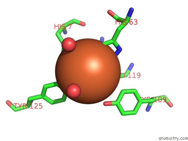
Mono view
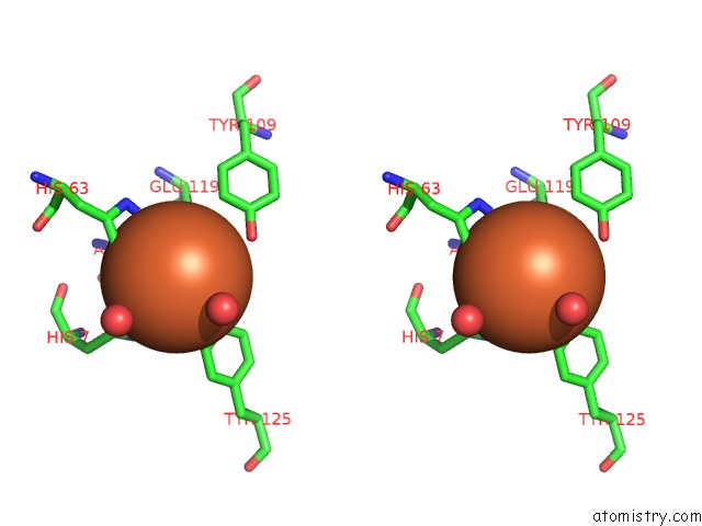
Stereo pair view
Mono view
Stereo pair view
A full contact list of Iron with other atoms in the Fe binding
site number 1 of Oxidase Chap within 5.0Å range:
|
Iron binding site 2 out of 2 in 6a4z
Go back to
Iron binding site 2 out
of 2 in the Oxidase Chap
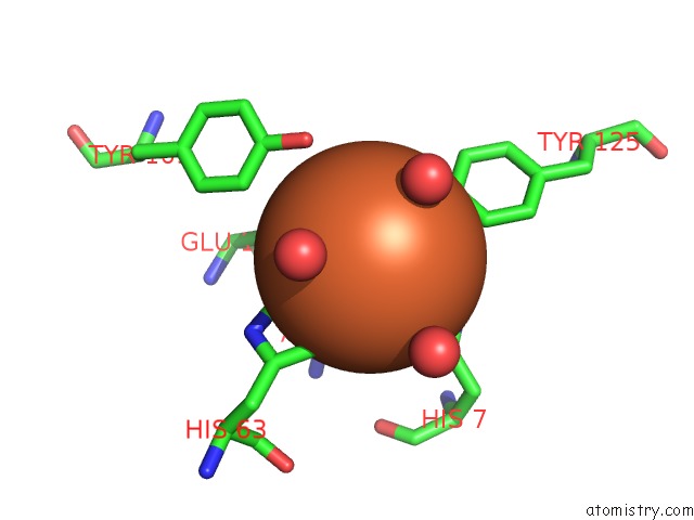
Mono view
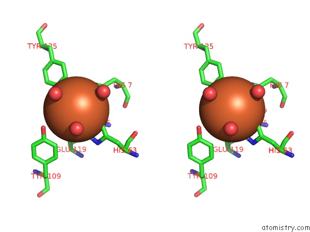
Stereo pair view
Mono view
Stereo pair view
A full contact list of Iron with other atoms in the Fe binding
site number 2 of Oxidase Chap within 5.0Å range:
|
Reference:
Y.S.Wang,
B.Zhang,
J.Zhu,
C.L.Yang,
Y.Guo,
C.L.Liu,
F.Liu,
H.Huang,
S.Zhao,
Y.Liang,
R.H.Jiao,
R.X.Tan,
H.M.Ge.
Molecular Basis For the Final Oxidative Rearrangement Steps in Chartreusin Biosynthesis. J. Am. Chem. Soc. V. 140 10909 2018.
ISSN: ESSN 1520-5126
PubMed: 30067334
DOI: 10.1021/JACS.8B06623
Page generated: Tue Aug 6 13:27:06 2024
ISSN: ESSN 1520-5126
PubMed: 30067334
DOI: 10.1021/JACS.8B06623
Last articles
F in 7LI5F in 7LHZ
F in 7LH7
F in 7LDE
F in 7LEP
F in 7LDD
F in 7LGX
F in 7LGK
F in 7LG8
F in 7LD3