Iron »
PDB 5zm9-6air »
6a7i »
Iron in PDB 6a7i: CYP154C4 From Streptomyces Sp. W2061
Protein crystallography data
The structure of CYP154C4 From Streptomyces Sp. W2061, PDB code: 6a7i
was solved by
C.W.Lee,
J.H.Lee,
with X-Ray Crystallography technique. A brief refinement statistics is given in the table below:
| Resolution Low / High (Å) | 46.47 / 2.19 |
| Space group | I 21 21 21 |
| Cell size a, b, c (Å), α, β, γ (°) | 83.755, 115.032, 127.807, 90.00, 90.00, 90.00 |
| R / Rfree (%) | 17.3 / 22.2 |
Iron Binding Sites:
The binding sites of Iron atom in the CYP154C4 From Streptomyces Sp. W2061
(pdb code 6a7i). This binding sites where shown within
5.0 Angstroms radius around Iron atom.
In total only one binding site of Iron was determined in the CYP154C4 From Streptomyces Sp. W2061, PDB code: 6a7i:
In total only one binding site of Iron was determined in the CYP154C4 From Streptomyces Sp. W2061, PDB code: 6a7i:
Iron binding site 1 out of 1 in 6a7i
Go back to
Iron binding site 1 out
of 1 in the CYP154C4 From Streptomyces Sp. W2061
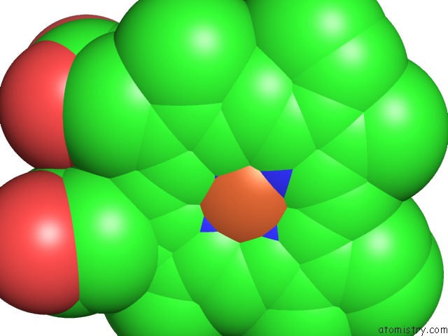
Mono view
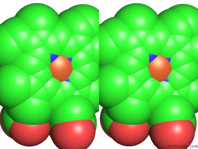
Stereo pair view
Mono view
Stereo pair view
A full contact list of Iron with other atoms in the Fe binding
site number 1 of CYP154C4 From Streptomyces Sp. W2061 within 5.0Å range:
|
Reference:
B.Dangi,
C.W.Lee,
K.H.Kim,
S.H.Park,
E.J.Yu,
C.S.Jeong,
H.Park,
J.H.Lee,
T.J.Oh.
Characterization of Two Steroid Hydroxylases From Different Streptomyces Spp. and Their Ligand-Bound and -Unbound Crystal Structures. Febs J. V. 286 1683 2019.
ISSN: ISSN 1742-464X
PubMed: 30552795
DOI: 10.1111/FEBS.14729
Page generated: Tue Aug 6 13:28:37 2024
ISSN: ISSN 1742-464X
PubMed: 30552795
DOI: 10.1111/FEBS.14729
Last articles
Cl in 5TR9Cl in 5TQU
Cl in 5TQJ
Cl in 5TPI
Cl in 5TQI
Cl in 5TPU
Cl in 5TPH
Cl in 5TPX
Cl in 5TPG
Cl in 5TOW