Iron »
PDB 5zm9-6air »
6acn »
Iron in PDB 6acn: Structure of Activated Aconitase. Formation of the (4FE-4S) Cluster in the Crystal
Enzymatic activity of Structure of Activated Aconitase. Formation of the (4FE-4S) Cluster in the Crystal
All present enzymatic activity of Structure of Activated Aconitase. Formation of the (4FE-4S) Cluster in the Crystal:
4.2.1.3;
4.2.1.3;
Protein crystallography data
The structure of Structure of Activated Aconitase. Formation of the (4FE-4S) Cluster in the Crystal, PDB code: 6acn
was solved by
A.H.Robbins,
C.D.Stout,
with X-Ray Crystallography technique. A brief refinement statistics is given in the table below:
| Resolution Low / High (Å) | 5.00 / 2.50 |
| Space group | P 21 21 2 |
| Cell size a, b, c (Å), α, β, γ (°) | 173.600, 72.000, 72.700, 90.00, 90.00, 90.00 |
| R / Rfree (%) | 18.7 / n/a |
Iron Binding Sites:
The binding sites of Iron atom in the Structure of Activated Aconitase. Formation of the (4FE-4S) Cluster in the Crystal
(pdb code 6acn). This binding sites where shown within
5.0 Angstroms radius around Iron atom.
In total 4 binding sites of Iron where determined in the Structure of Activated Aconitase. Formation of the (4FE-4S) Cluster in the Crystal, PDB code: 6acn:
Jump to Iron binding site number: 1; 2; 3; 4;
In total 4 binding sites of Iron where determined in the Structure of Activated Aconitase. Formation of the (4FE-4S) Cluster in the Crystal, PDB code: 6acn:
Jump to Iron binding site number: 1; 2; 3; 4;
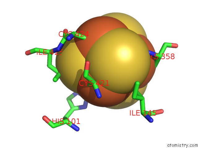
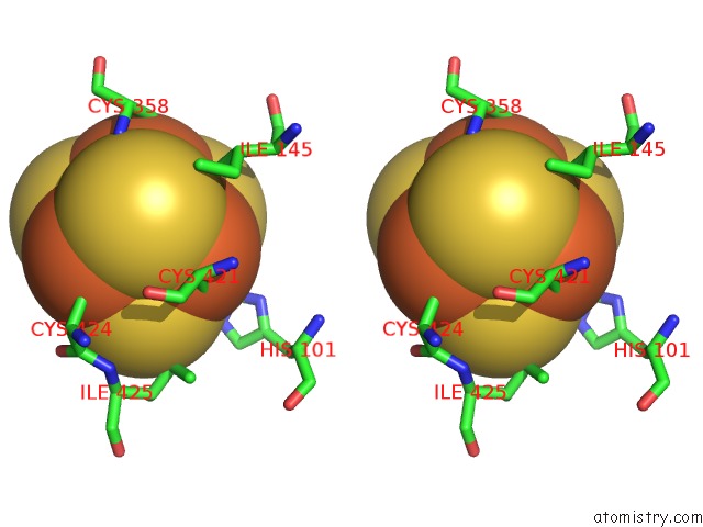
Iron binding site 1 out of 4 in 6acn
Go back to
Iron binding site 1 out
of 4 in the Structure of Activated Aconitase. Formation of the (4FE-4S) Cluster in the Crystal
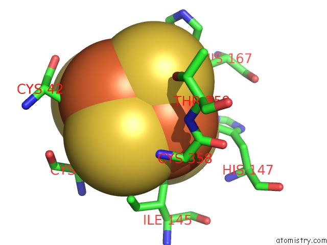
Mono view
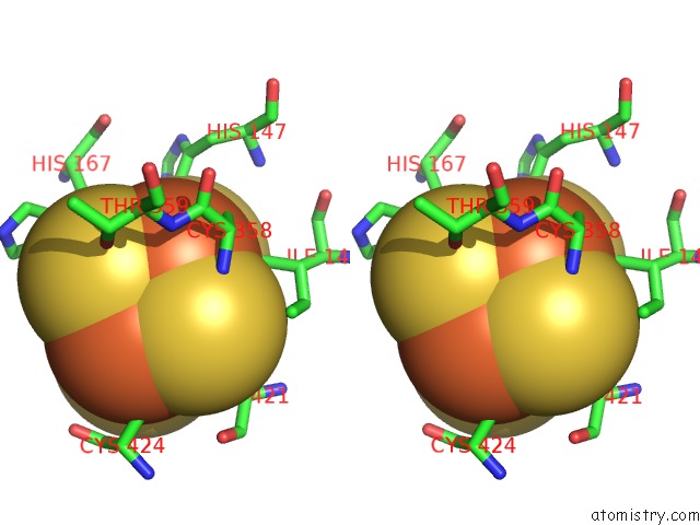
Stereo pair view
Mono view
Stereo pair view
A full contact list of Iron with other atoms in the Fe binding
site number 1 of Structure of Activated Aconitase. Formation of the (4FE-4S) Cluster in the Crystal within 5.0Å range:
|
Iron binding site 2 out of 4 in 6acn
Go back to
Iron binding site 2 out
of 4 in the Structure of Activated Aconitase. Formation of the (4FE-4S) Cluster in the Crystal
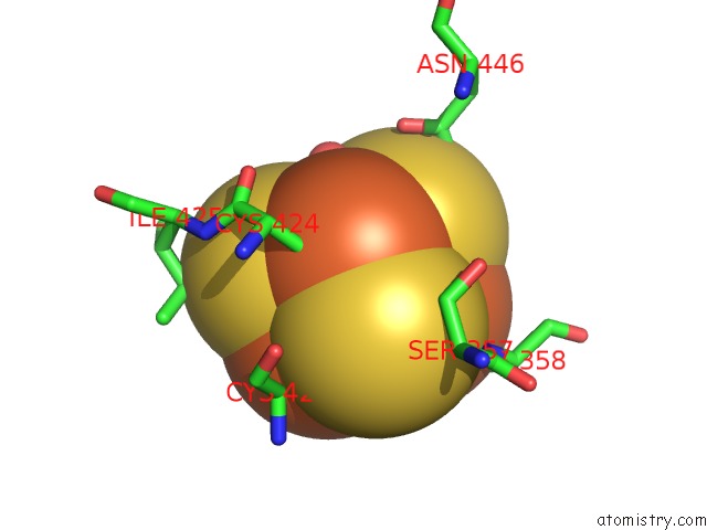
Mono view
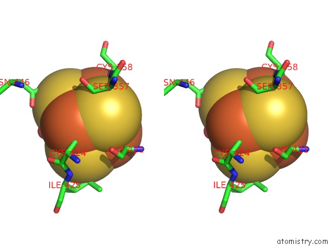
Stereo pair view
Mono view
Stereo pair view
A full contact list of Iron with other atoms in the Fe binding
site number 2 of Structure of Activated Aconitase. Formation of the (4FE-4S) Cluster in the Crystal within 5.0Å range:
|
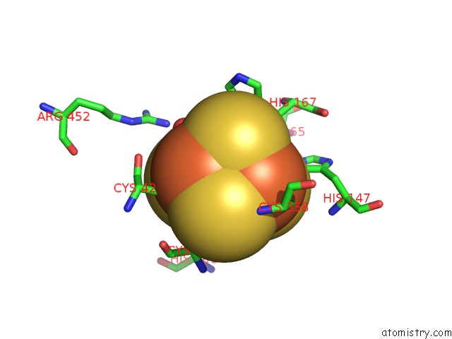
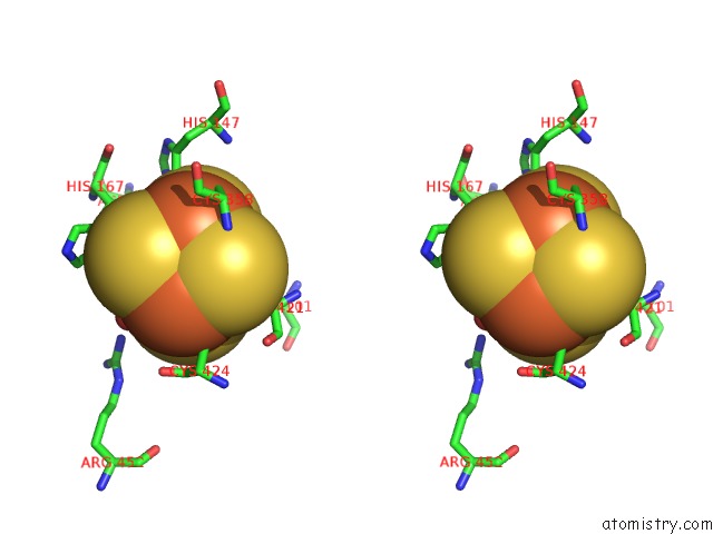
Iron binding site 3 out of 4 in 6acn
Go back to
Iron binding site 3 out
of 4 in the Structure of Activated Aconitase. Formation of the (4FE-4S) Cluster in the Crystal
Mono view
Stereo pair view
Mono view
Stereo pair view
A full contact list of Iron with other atoms in the Fe binding
site number 3 of Structure of Activated Aconitase. Formation of the (4FE-4S) Cluster in the Crystal within 5.0Å range:
|
Iron binding site 4 out of 4 in 6acn
Go back to
Iron binding site 4 out
of 4 in the Structure of Activated Aconitase. Formation of the (4FE-4S) Cluster in the Crystal
Mono view
Stereo pair view
Mono view
Stereo pair view
A full contact list of Iron with other atoms in the Fe binding
site number 4 of Structure of Activated Aconitase. Formation of the (4FE-4S) Cluster in the Crystal within 5.0Å range:
|
Reference:
A.H.Robbins,
C.D.Stout.
Structure of Activated Aconitase: Formation of the [4FE-4S] Cluster in the Crystal. Proc.Natl.Acad.Sci.Usa V. 86 3639 1989.
ISSN: ISSN 0027-8424
PubMed: 2726740
DOI: 10.1073/PNAS.86.10.3639
Page generated: Tue Aug 6 13:30:10 2024
ISSN: ISSN 0027-8424
PubMed: 2726740
DOI: 10.1073/PNAS.86.10.3639
Last articles
Fe in 2YXOFe in 2YRS
Fe in 2YXC
Fe in 2YNM
Fe in 2YVJ
Fe in 2YP1
Fe in 2YU2
Fe in 2YU1
Fe in 2YQB
Fe in 2YOO