Iron »
PDB 6ajo-6ayc »
6ajr »
Iron in PDB 6ajr: Complex Form of Uracil Dna Glycosylase X and Uracil
Protein crystallography data
The structure of Complex Form of Uracil Dna Glycosylase X and Uracil, PDB code: 6ajr
was solved by
W.C.Ahn,
S.Aroli,
U.Varshney,
E.J.Woo,
with X-Ray Crystallography technique. A brief refinement statistics is given in the table below:
| Resolution Low / High (Å) | 19.01 / 1.34 |
| Space group | P 1 21 1 |
| Cell size a, b, c (Å), α, β, γ (°) | 36.479, 51.773, 55.107, 90.00, 104.98, 90.00 |
| R / Rfree (%) | 13.8 / 16.6 |
Iron Binding Sites:
The binding sites of Iron atom in the Complex Form of Uracil Dna Glycosylase X and Uracil
(pdb code 6ajr). This binding sites where shown within
5.0 Angstroms radius around Iron atom.
In total 4 binding sites of Iron where determined in the Complex Form of Uracil Dna Glycosylase X and Uracil, PDB code: 6ajr:
Jump to Iron binding site number: 1; 2; 3; 4;
In total 4 binding sites of Iron where determined in the Complex Form of Uracil Dna Glycosylase X and Uracil, PDB code: 6ajr:
Jump to Iron binding site number: 1; 2; 3; 4;
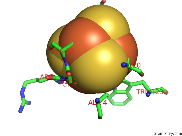
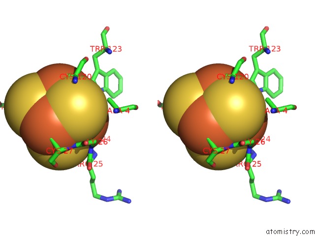
Iron binding site 1 out of 4 in 6ajr
Go back to
Iron binding site 1 out
of 4 in the Complex Form of Uracil Dna Glycosylase X and Uracil
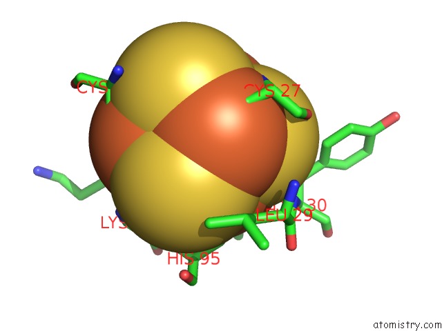
Mono view
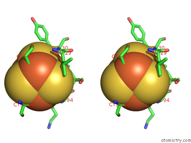
Stereo pair view
Mono view
Stereo pair view
A full contact list of Iron with other atoms in the Fe binding
site number 1 of Complex Form of Uracil Dna Glycosylase X and Uracil within 5.0Å range:
|
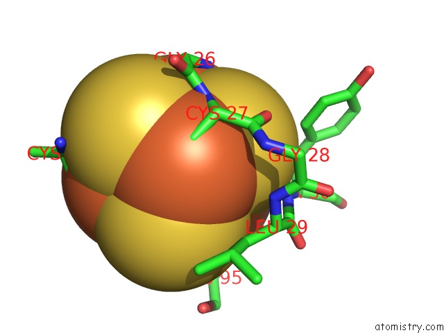
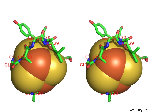
Iron binding site 2 out of 4 in 6ajr
Go back to
Iron binding site 2 out
of 4 in the Complex Form of Uracil Dna Glycosylase X and Uracil
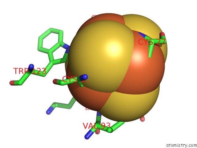
Mono view
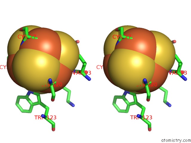
Stereo pair view
Mono view
Stereo pair view
A full contact list of Iron with other atoms in the Fe binding
site number 2 of Complex Form of Uracil Dna Glycosylase X and Uracil within 5.0Å range:
|
Iron binding site 3 out of 4 in 6ajr
Go back to
Iron binding site 3 out
of 4 in the Complex Form of Uracil Dna Glycosylase X and Uracil
Mono view
Stereo pair view
Mono view
Stereo pair view
A full contact list of Iron with other atoms in the Fe binding
site number 3 of Complex Form of Uracil Dna Glycosylase X and Uracil within 5.0Å range:
|
Iron binding site 4 out of 4 in 6ajr
Go back to
Iron binding site 4 out
of 4 in the Complex Form of Uracil Dna Glycosylase X and Uracil
Mono view
Stereo pair view
Mono view
Stereo pair view
A full contact list of Iron with other atoms in the Fe binding
site number 4 of Complex Form of Uracil Dna Glycosylase X and Uracil within 5.0Å range:
|
Reference:
W.C.Ahn,
S.Aroli,
J.H.Kim,
J.H.Moon,
G.S.Lee,
M.H.Lee,
P.B.Sang,
B.H.Oh,
U.Varshney,
E.J.Woo.
Covalent Binding of Uracil Dna Glycosylase Udgx to Abasic Dna Upon Uracil Excision. Nat.Chem.Biol. V. 15 607 2019.
ISSN: ESSN 1552-4469
PubMed: 31101917
DOI: 10.1038/S41589-019-0289-3
Page generated: Tue Aug 6 13:45:08 2024
ISSN: ESSN 1552-4469
PubMed: 31101917
DOI: 10.1038/S41589-019-0289-3
Last articles
F in 7O7KF in 7OFA
F in 7OAM
F in 7O7J
F in 7O75
F in 7O73
F in 7O72
F in 7O70
F in 7O6I
F in 7O4J