Iron »
PDB 6ajo-6ayc »
6ajs »
Iron in PDB 6ajs: H109S Mutant Form of Uracil Dna Glycosylase X.
Protein crystallography data
The structure of H109S Mutant Form of Uracil Dna Glycosylase X., PDB code: 6ajs
was solved by
W.C.Ahn,
S.Aroli,
U.Varshney,
E.J.Woo,
with X-Ray Crystallography technique. A brief refinement statistics is given in the table below:
| Resolution Low / High (Å) | 28.02 / 1.63 |
| Space group | P 1 21 1 |
| Cell size a, b, c (Å), α, β, γ (°) | 36.229, 51.393, 54.444, 90.00, 105.21, 90.00 |
| R / Rfree (%) | 16 / 19.7 |
Iron Binding Sites:
The binding sites of Iron atom in the H109S Mutant Form of Uracil Dna Glycosylase X.
(pdb code 6ajs). This binding sites where shown within
5.0 Angstroms radius around Iron atom.
In total 4 binding sites of Iron where determined in the H109S Mutant Form of Uracil Dna Glycosylase X., PDB code: 6ajs:
Jump to Iron binding site number: 1; 2; 3; 4;
In total 4 binding sites of Iron where determined in the H109S Mutant Form of Uracil Dna Glycosylase X., PDB code: 6ajs:
Jump to Iron binding site number: 1; 2; 3; 4;
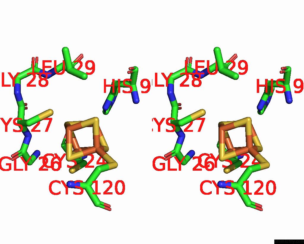
Iron binding site 1 out of 4 in 6ajs
Go back to
Iron binding site 1 out
of 4 in the H109S Mutant Form of Uracil Dna Glycosylase X.

Mono view

Stereo pair view
Mono view
Stereo pair view
A full contact list of Iron with other atoms in the Fe binding
site number 1 of H109S Mutant Form of Uracil Dna Glycosylase X. within 5.0Å range:
|
Iron binding site 2 out of 4 in 6ajs
Go back to
Iron binding site 2 out
of 4 in the H109S Mutant Form of Uracil Dna Glycosylase X.

Mono view
Stereo pair view
Mono view
Stereo pair view
A full contact list of Iron with other atoms in the Fe binding
site number 2 of H109S Mutant Form of Uracil Dna Glycosylase X. within 5.0Å range:
|
Iron binding site 3 out of 4 in 6ajs
Go back to
Iron binding site 3 out
of 4 in the H109S Mutant Form of Uracil Dna Glycosylase X.

Mono view

Stereo pair view
Mono view
Stereo pair view
A full contact list of Iron with other atoms in the Fe binding
site number 3 of H109S Mutant Form of Uracil Dna Glycosylase X. within 5.0Å range:
|
Iron binding site 4 out of 4 in 6ajs
Go back to
Iron binding site 4 out
of 4 in the H109S Mutant Form of Uracil Dna Glycosylase X.

Mono view

Stereo pair view
Mono view
Stereo pair view
A full contact list of Iron with other atoms in the Fe binding
site number 4 of H109S Mutant Form of Uracil Dna Glycosylase X. within 5.0Å range:
|
Reference:
W.C.Ahn,
S.Aroli,
J.H.Kim,
J.H.Moon,
G.S.Lee,
M.H.Lee,
P.B.Sang,
B.H.Oh,
U.Varshney,
E.J.Woo.
Covalent Binding of Uracil Dna Glycosylase Udgx to Abasic Dna Upon Uracil Excision. Nat.Chem.Biol. V. 15 607 2019.
ISSN: ESSN 1552-4469
PubMed: 31101917
DOI: 10.1038/S41589-019-0289-3
Page generated: Tue Aug 6 13:45:08 2024
ISSN: ESSN 1552-4469
PubMed: 31101917
DOI: 10.1038/S41589-019-0289-3
Last articles
Zn in 9JYWZn in 9IR4
Zn in 9IR3
Zn in 9GMX
Zn in 9GMW
Zn in 9JEJ
Zn in 9ERF
Zn in 9ERE
Zn in 9EGV
Zn in 9EGW