Iron »
PDB 6ajo-6ayc »
6alo »
Iron in PDB 6alo: Vioc L-Arginine Hydroxylase Bound to Fe(II), L-Arginine, and A Peroxysuccinate Intermediate
Enzymatic activity of Vioc L-Arginine Hydroxylase Bound to Fe(II), L-Arginine, and A Peroxysuccinate Intermediate
All present enzymatic activity of Vioc L-Arginine Hydroxylase Bound to Fe(II), L-Arginine, and A Peroxysuccinate Intermediate:
1.14.11.41;
1.14.11.41;
Protein crystallography data
The structure of Vioc L-Arginine Hydroxylase Bound to Fe(II), L-Arginine, and A Peroxysuccinate Intermediate, PDB code: 6alo
was solved by
N.P.Dunham,
A.J.Mitchell,
A.K.Boal,
with X-Ray Crystallography technique. A brief refinement statistics is given in the table below:
| Resolution Low / High (Å) | 59.39 / 1.79 |
| Space group | C 1 2 1 |
| Cell size a, b, c (Å), α, β, γ (°) | 80.760, 66.653, 62.889, 90.00, 109.21, 90.00 |
| R / Rfree (%) | 17.5 / 20.3 |
Iron Binding Sites:
The binding sites of Iron atom in the Vioc L-Arginine Hydroxylase Bound to Fe(II), L-Arginine, and A Peroxysuccinate Intermediate
(pdb code 6alo). This binding sites where shown within
5.0 Angstroms radius around Iron atom.
In total only one binding site of Iron was determined in the Vioc L-Arginine Hydroxylase Bound to Fe(II), L-Arginine, and A Peroxysuccinate Intermediate, PDB code: 6alo:
In total only one binding site of Iron was determined in the Vioc L-Arginine Hydroxylase Bound to Fe(II), L-Arginine, and A Peroxysuccinate Intermediate, PDB code: 6alo:
Iron binding site 1 out of 1 in 6alo
Go back to
Iron binding site 1 out
of 1 in the Vioc L-Arginine Hydroxylase Bound to Fe(II), L-Arginine, and A Peroxysuccinate Intermediate
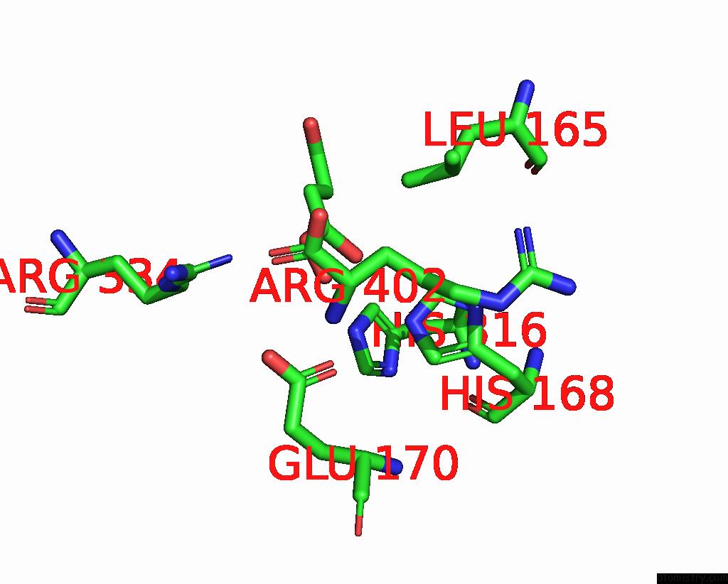
Mono view
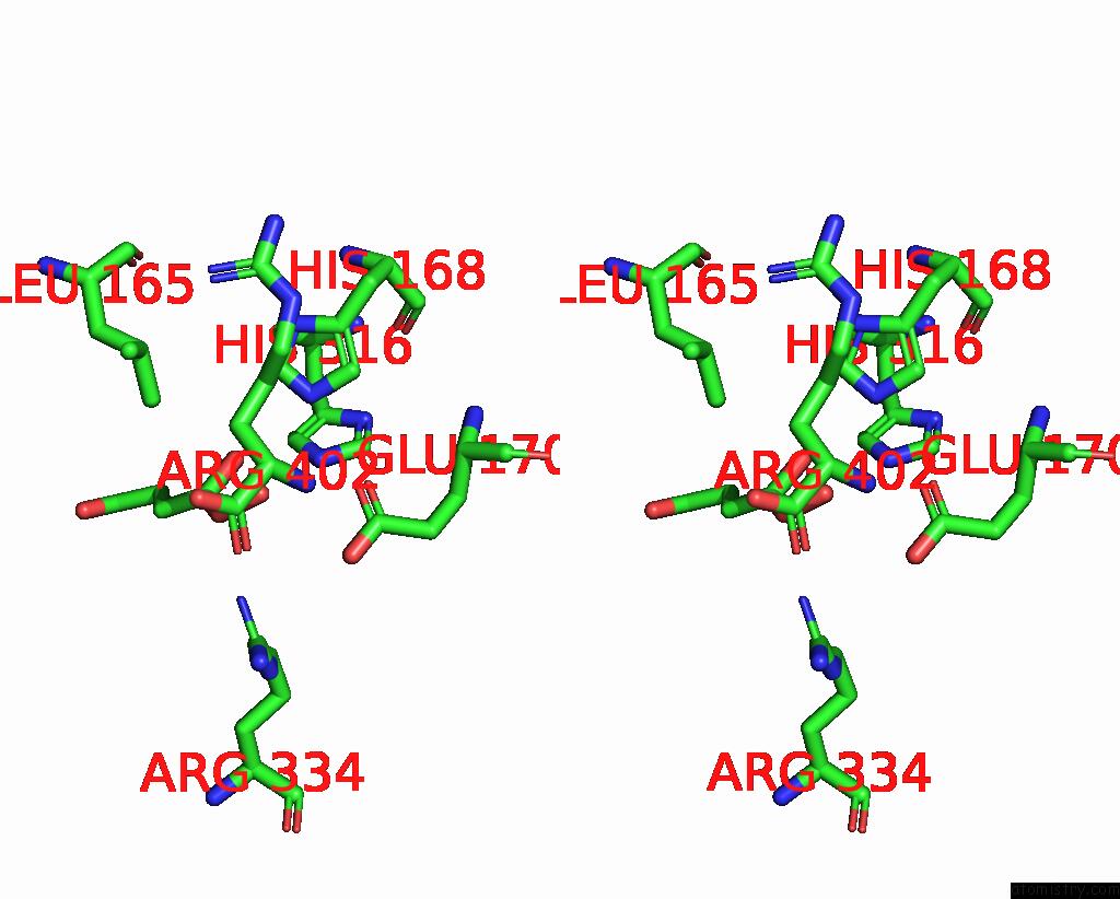
Stereo pair view
Mono view
Stereo pair view
A full contact list of Iron with other atoms in the Fe binding
site number 1 of Vioc L-Arginine Hydroxylase Bound to Fe(II), L-Arginine, and A Peroxysuccinate Intermediate within 5.0Å range:
|
Reference:
A.J.Mitchell,
N.P.Dunham,
R.J.Martinie,
J.A.Bergman,
C.J.Pollock,
K.Hu,
B.D.Allen,
W.C.Chang,
A.Silakov,
J.M.Bollinger,
C.Krebs,
A.K.Boal.
Visualizing the Reaction Cycle in An Iron(II)- and 2-(Oxo)-Glutarate-Dependent Hydroxylase. J. Am. Chem. Soc. V. 139 13830 2017.
ISSN: ESSN 1520-5126
PubMed: 28823155
DOI: 10.1021/JACS.7B07374
Page generated: Tue Aug 6 13:46:20 2024
ISSN: ESSN 1520-5126
PubMed: 28823155
DOI: 10.1021/JACS.7B07374
Last articles
Cl in 8BZLCl in 8C3L
Cl in 8C2F
Cl in 8C25
Cl in 8C3F
Cl in 8C2E
Cl in 8C1P
Cl in 8C26
Cl in 8C1K
Cl in 8C1H