Iron »
PDB 6azu-6bpt »
6bdj »
Iron in PDB 6bdj: Crystal Structure of Dioxygenase TETUR07G02040
Protein crystallography data
The structure of Crystal Structure of Dioxygenase TETUR07G02040, PDB code: 6bdj
was solved by
L.Daneshian,
C.R.Schlachter,
M.Chruszcz,
with X-Ray Crystallography technique. A brief refinement statistics is given in the table below:
| Resolution Low / High (Å) | 40.00 / 2.15 |
| Space group | P 1 2 1 |
| Cell size a, b, c (Å), α, β, γ (°) | 61.091, 45.415, 83.040, 90.00, 94.29, 90.00 |
| R / Rfree (%) | 21.4 / 24.8 |
Iron Binding Sites:
The binding sites of Iron atom in the Crystal Structure of Dioxygenase TETUR07G02040
(pdb code 6bdj). This binding sites where shown within
5.0 Angstroms radius around Iron atom.
In total 2 binding sites of Iron where determined in the Crystal Structure of Dioxygenase TETUR07G02040, PDB code: 6bdj:
Jump to Iron binding site number: 1; 2;
In total 2 binding sites of Iron where determined in the Crystal Structure of Dioxygenase TETUR07G02040, PDB code: 6bdj:
Jump to Iron binding site number: 1; 2;
Iron binding site 1 out of 2 in 6bdj
Go back to
Iron binding site 1 out
of 2 in the Crystal Structure of Dioxygenase TETUR07G02040
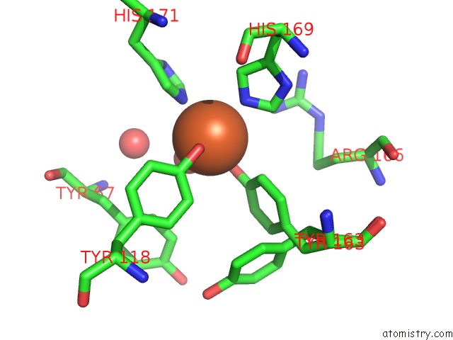
Mono view
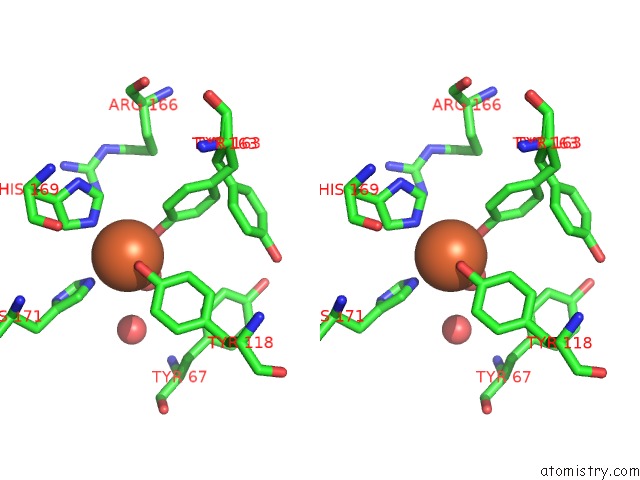
Stereo pair view
Mono view
Stereo pair view
A full contact list of Iron with other atoms in the Fe binding
site number 1 of Crystal Structure of Dioxygenase TETUR07G02040 within 5.0Å range:
|
Iron binding site 2 out of 2 in 6bdj
Go back to
Iron binding site 2 out
of 2 in the Crystal Structure of Dioxygenase TETUR07G02040
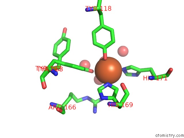
Mono view
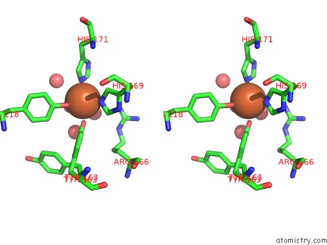
Stereo pair view
Mono view
Stereo pair view
A full contact list of Iron with other atoms in the Fe binding
site number 2 of Crystal Structure of Dioxygenase TETUR07G02040 within 5.0Å range:
|
Reference:
C.R.Schlachter,
L.Daneshian,
J.Amaya,
V.Klapper,
N.Wybouw,
T.Borowski,
T.Van Leeuwen,
V.Grbic,
M.Grbic,
T.M.Makris,
M.Chruszcz.
Structural and Functional Characterization of An Intradiol Ring-Cleavage Dioxygenase From the Polyphagous Spider Mite Herbivore Tetranychus Urticae Koch. Insect Biochem.Mol.Biol. V. 107 19 2019.
ISSN: ISSN 0965-1748
PubMed: 30529144
DOI: 10.1016/J.IBMB.2018.12.001
Page generated: Tue Aug 6 14:15:13 2024
ISSN: ISSN 0965-1748
PubMed: 30529144
DOI: 10.1016/J.IBMB.2018.12.001
Last articles
Zn in 9J0NZn in 9J0O
Zn in 9J0P
Zn in 9FJX
Zn in 9EKB
Zn in 9C0F
Zn in 9CAH
Zn in 9CH0
Zn in 9CH3
Zn in 9CH1