Iron »
PDB 6bpu-6cdh »
6bww »
Iron in PDB 6bww: Crystal Structure of An Acetate and Cymal-5 Bound Cytochrome P450 2B4 F429H Mutant
Enzymatic activity of Crystal Structure of An Acetate and Cymal-5 Bound Cytochrome P450 2B4 F429H Mutant
All present enzymatic activity of Crystal Structure of An Acetate and Cymal-5 Bound Cytochrome P450 2B4 F429H Mutant:
1.14.14.1;
1.14.14.1;
Protein crystallography data
The structure of Crystal Structure of An Acetate and Cymal-5 Bound Cytochrome P450 2B4 F429H Mutant, PDB code: 6bww
was solved by
Y.T.Yang,
L.Waskell,
with X-Ray Crystallography technique. A brief refinement statistics is given in the table below:
| Resolution Low / High (Å) | 46.59 / 2.10 |
| Space group | P 31 2 1 |
| Cell size a, b, c (Å), α, β, γ (°) | 93.185, 93.185, 151.092, 90.00, 90.00, 120.00 |
| R / Rfree (%) | 18.5 / 20.6 |
Iron Binding Sites:
The binding sites of Iron atom in the Crystal Structure of An Acetate and Cymal-5 Bound Cytochrome P450 2B4 F429H Mutant
(pdb code 6bww). This binding sites where shown within
5.0 Angstroms radius around Iron atom.
In total only one binding site of Iron was determined in the Crystal Structure of An Acetate and Cymal-5 Bound Cytochrome P450 2B4 F429H Mutant, PDB code: 6bww:
In total only one binding site of Iron was determined in the Crystal Structure of An Acetate and Cymal-5 Bound Cytochrome P450 2B4 F429H Mutant, PDB code: 6bww:
Iron binding site 1 out of 1 in 6bww
Go back to
Iron binding site 1 out
of 1 in the Crystal Structure of An Acetate and Cymal-5 Bound Cytochrome P450 2B4 F429H Mutant
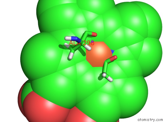
Mono view
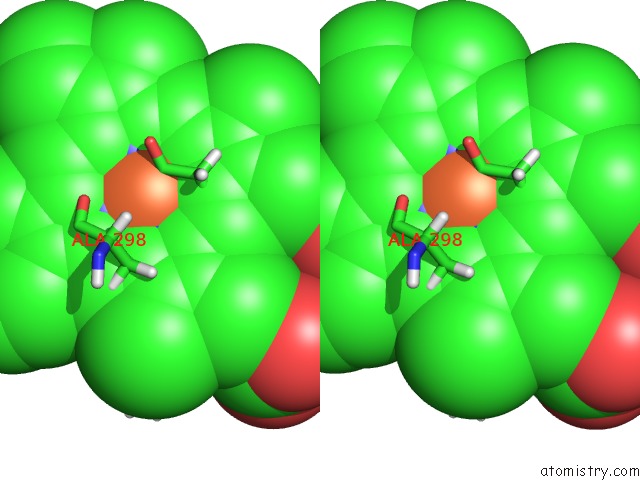
Stereo pair view
Mono view
Stereo pair view
A full contact list of Iron with other atoms in the Fe binding
site number 1 of Crystal Structure of An Acetate and Cymal-5 Bound Cytochrome P450 2B4 F429H Mutant within 5.0Å range:
|
Reference:
Y.Yang,
W.Bu,
S.Im,
J.Meagher,
J.Stuckey,
L.Waskell.
Structure of Cytochrome P450 2B4 with An Acetate Ligand and An Active Site Hydrogen Bond Network Similar to Oxyferrous P450CAM. J.Inorg.Biochem. V. 185 17 2018.
ISSN: ISSN 0162-0134
PubMed: 29730233
DOI: 10.1016/J.JINORGBIO.2018.04.015
Page generated: Tue Aug 6 14:39:05 2024
ISSN: ISSN 0162-0134
PubMed: 29730233
DOI: 10.1016/J.JINORGBIO.2018.04.015
Last articles
Zn in 9J0NZn in 9J0O
Zn in 9J0P
Zn in 9FJX
Zn in 9EKB
Zn in 9C0F
Zn in 9CAH
Zn in 9CH0
Zn in 9CH3
Zn in 9CH1