Iron »
PDB 6cdk-6cxu »
6cl6 »
Iron in PDB 6cl6: Structure of P. Aeruginosa R2 Pyocin Fiber PA0620 Comprising C- Terminal Residues 323-691
Protein crystallography data
The structure of Structure of P. Aeruginosa R2 Pyocin Fiber PA0620 Comprising C- Terminal Residues 323-691, PDB code: 6cl6
was solved by
S.A.Buth,
M.M.Shneider,
P.G.Leiman,
with X-Ray Crystallography technique. A brief refinement statistics is given in the table below:
| Resolution Low / High (Å) | 49.96 / 1.90 |
| Space group | P 21 21 21 |
| Cell size a, b, c (Å), α, β, γ (°) | 56.320, 126.946, 432.938, 90.00, 90.00, 90.00 |
| R / Rfree (%) | 16 / 20.8 |
Other elements in 6cl6:
The structure of Structure of P. Aeruginosa R2 Pyocin Fiber PA0620 Comprising C- Terminal Residues 323-691 also contains other interesting chemical elements:
| Magnesium | (Mg) | 2 atoms |
| Calcium | (Ca) | 6 atoms |
Iron Binding Sites:
The binding sites of Iron atom in the Structure of P. Aeruginosa R2 Pyocin Fiber PA0620 Comprising C- Terminal Residues 323-691
(pdb code 6cl6). This binding sites where shown within
5.0 Angstroms radius around Iron atom.
In total 2 binding sites of Iron where determined in the Structure of P. Aeruginosa R2 Pyocin Fiber PA0620 Comprising C- Terminal Residues 323-691, PDB code: 6cl6:
Jump to Iron binding site number: 1; 2;
In total 2 binding sites of Iron where determined in the Structure of P. Aeruginosa R2 Pyocin Fiber PA0620 Comprising C- Terminal Residues 323-691, PDB code: 6cl6:
Jump to Iron binding site number: 1; 2;
Iron binding site 1 out of 2 in 6cl6
Go back to
Iron binding site 1 out
of 2 in the Structure of P. Aeruginosa R2 Pyocin Fiber PA0620 Comprising C- Terminal Residues 323-691
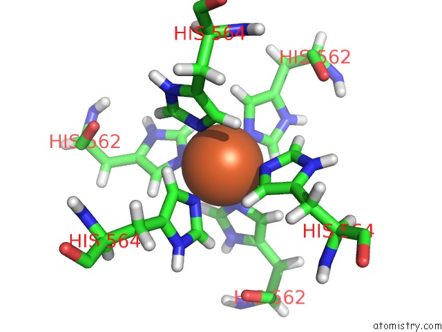
Mono view
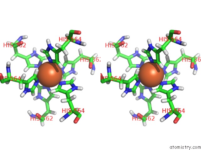
Stereo pair view
Mono view
Stereo pair view
A full contact list of Iron with other atoms in the Fe binding
site number 1 of Structure of P. Aeruginosa R2 Pyocin Fiber PA0620 Comprising C- Terminal Residues 323-691 within 5.0Å range:
|
Iron binding site 2 out of 2 in 6cl6
Go back to
Iron binding site 2 out
of 2 in the Structure of P. Aeruginosa R2 Pyocin Fiber PA0620 Comprising C- Terminal Residues 323-691
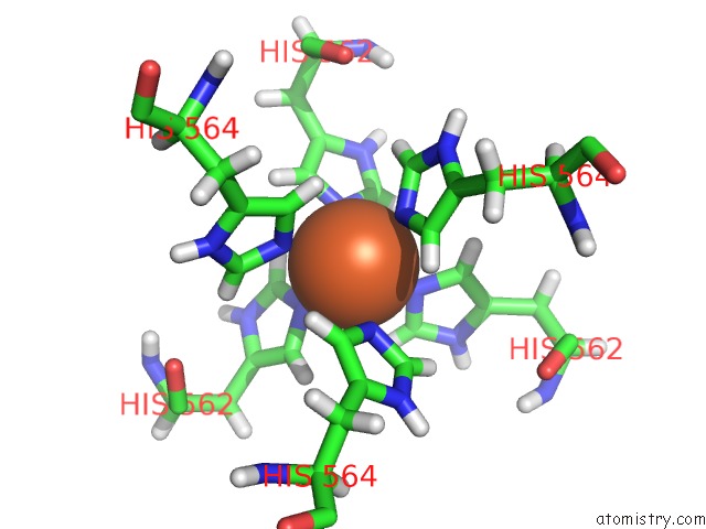
Mono view
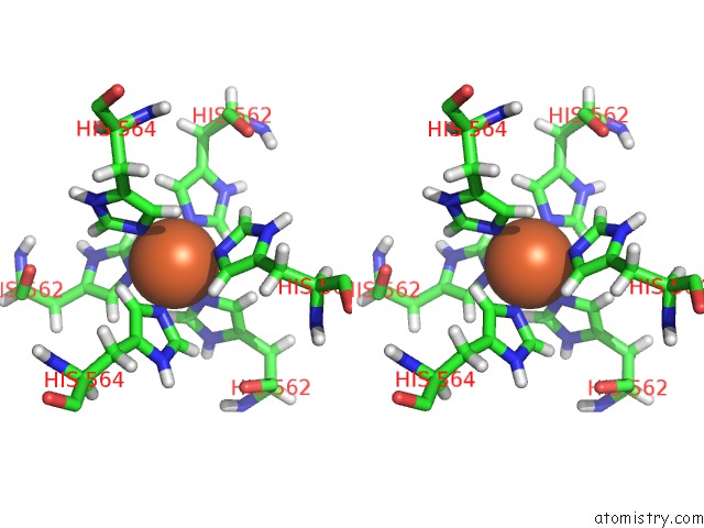
Stereo pair view
Mono view
Stereo pair view
A full contact list of Iron with other atoms in the Fe binding
site number 2 of Structure of P. Aeruginosa R2 Pyocin Fiber PA0620 Comprising C- Terminal Residues 323-691 within 5.0Å range:
|
Reference:
S.A.Buth,
M.M.Shneider,
D.Scholl,
P.G.Leiman.
Structure and Analysis of R1 and R2 Pyocin Receptor-Binding Fibers. Viruses V. 10 2018.
ISSN: ISSN 1999-4915
PubMed: 30110933
DOI: 10.3390/V10080427
Page generated: Wed Aug 6 04:52:18 2025
ISSN: ISSN 1999-4915
PubMed: 30110933
DOI: 10.3390/V10080427
Last articles
Fe in 6G7MFe in 6G74
Fe in 6G7Q
Fe in 6G7P
Fe in 6G7N
Fe in 6G71
Fe in 6G2J
Fe in 6G5Q
Fe in 6G5T
Fe in 6G5O