Iron »
PDB 6e45-6f0a »
6enk »
Iron in PDB 6enk: The X-Ray Crystal Structure of Dese Bound to Desferrioxamine B
Protein crystallography data
The structure of The X-Ray Crystal Structure of Dese Bound to Desferrioxamine B, PDB code: 6enk
was solved by
J.H.Naismith,
S.A.Mcmahon,
G.L.Challis,
N.Kadi,
M.Oke,
H.Liu,
L.G.Carter,
K.A.Johnson,
with X-Ray Crystallography technique. A brief refinement statistics is given in the table below:
| Resolution Low / High (Å) | 30.99 / 1.96 |
| Space group | P 21 21 21 |
| Cell size a, b, c (Å), α, β, γ (°) | 53.163, 58.055, 101.194, 90.00, 90.00, 90.00 |
| R / Rfree (%) | 15.9 / 20.4 |
Other elements in 6enk:
The structure of The X-Ray Crystal Structure of Dese Bound to Desferrioxamine B also contains other interesting chemical elements:
| Sodium | (Na) | 3 atoms |
Iron Binding Sites:
The binding sites of Iron atom in the The X-Ray Crystal Structure of Dese Bound to Desferrioxamine B
(pdb code 6enk). This binding sites where shown within
5.0 Angstroms radius around Iron atom.
In total only one binding site of Iron was determined in the The X-Ray Crystal Structure of Dese Bound to Desferrioxamine B, PDB code: 6enk:
In total only one binding site of Iron was determined in the The X-Ray Crystal Structure of Dese Bound to Desferrioxamine B, PDB code: 6enk:
Iron binding site 1 out of 1 in 6enk
Go back to
Iron binding site 1 out
of 1 in the The X-Ray Crystal Structure of Dese Bound to Desferrioxamine B
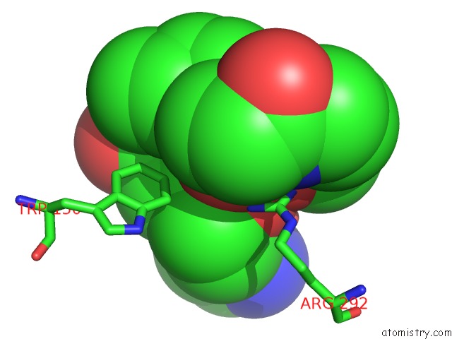
Mono view
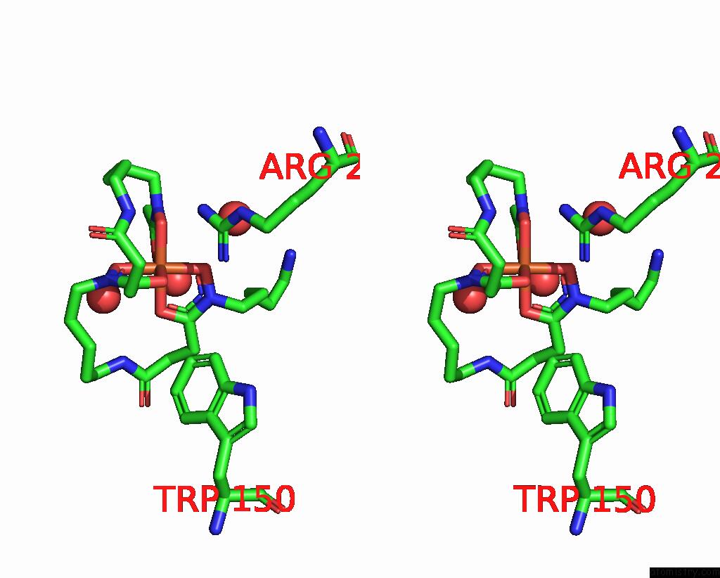
Stereo pair view
Mono view
Stereo pair view
A full contact list of Iron with other atoms in the Fe binding
site number 1 of The X-Ray Crystal Structure of Dese Bound to Desferrioxamine B within 5.0Å range:
|
Reference:
J.L.Ronan,
N.Kadi,
S.A.Mcmahon,
J.H.Naismith,
L.M.Alkhalaf,
G.L.Challis.
Desferrioxamine Biosynthesis: Diverse Hydroxamate Assembly By Substrate-Tolerant Acyl Transferase Desc. Philos. Trans. R. Soc. V. 373 2018LOND., B, Biol. Sci..
ISSN: ESSN 1471-2970
PubMed: 29685972
DOI: 10.1098/RSTB.2017.0068
Page generated: Tue Aug 6 17:27:24 2024
ISSN: ESSN 1471-2970
PubMed: 29685972
DOI: 10.1098/RSTB.2017.0068
Last articles
Zn in 9J0NZn in 9J0O
Zn in 9J0P
Zn in 9FJX
Zn in 9EKB
Zn in 9C0F
Zn in 9CAH
Zn in 9CH0
Zn in 9CH3
Zn in 9CH1