Iron »
PDB 6e45-6f0a »
6exf »
Iron in PDB 6exf: Crystal Structure of the Complex Fe(II)/Alpha-Ketoglutarate Dependent Dioxygenase KDO5 with Fe(II)/Lysine
Protein crystallography data
The structure of Crystal Structure of the Complex Fe(II)/Alpha-Ketoglutarate Dependent Dioxygenase KDO5 with Fe(II)/Lysine, PDB code: 6exf
was solved by
T.Isabet,
E.Stura,
P.Legrand,
A.Zaparucha,
K.Bastard,
with X-Ray Crystallography technique. A brief refinement statistics is given in the table below:
| Resolution Low / High (Å) | 28.55 / 1.95 |
| Space group | P 21 21 21 |
| Cell size a, b, c (Å), α, β, γ (°) | 91.721, 99.618, 165.819, 90.00, 90.00, 90.00 |
| R / Rfree (%) | 17.7 / 21.3 |
Iron Binding Sites:
The binding sites of Iron atom in the Crystal Structure of the Complex Fe(II)/Alpha-Ketoglutarate Dependent Dioxygenase KDO5 with Fe(II)/Lysine
(pdb code 6exf). This binding sites where shown within
5.0 Angstroms radius around Iron atom.
In total 4 binding sites of Iron where determined in the Crystal Structure of the Complex Fe(II)/Alpha-Ketoglutarate Dependent Dioxygenase KDO5 with Fe(II)/Lysine, PDB code: 6exf:
Jump to Iron binding site number: 1; 2; 3; 4;
In total 4 binding sites of Iron where determined in the Crystal Structure of the Complex Fe(II)/Alpha-Ketoglutarate Dependent Dioxygenase KDO5 with Fe(II)/Lysine, PDB code: 6exf:
Jump to Iron binding site number: 1; 2; 3; 4;
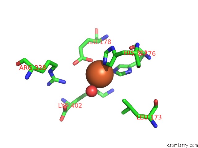
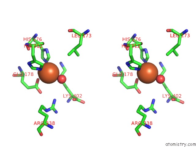
Iron binding site 1 out of 4 in 6exf
Go back to
Iron binding site 1 out
of 4 in the Crystal Structure of the Complex Fe(II)/Alpha-Ketoglutarate Dependent Dioxygenase KDO5 with Fe(II)/Lysine
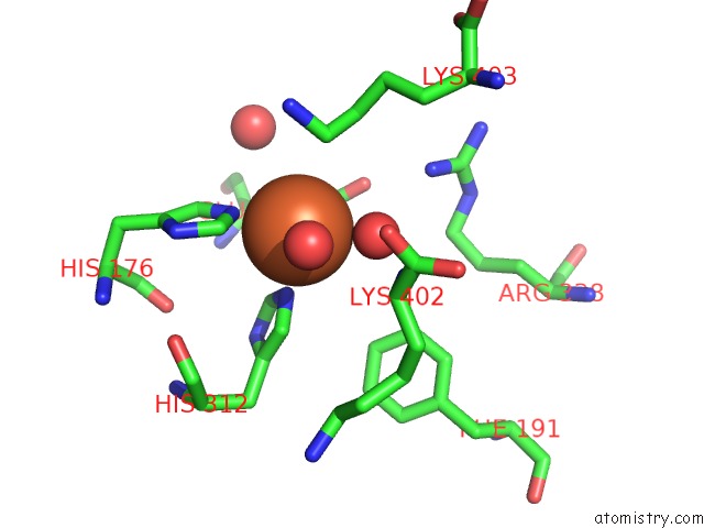
Mono view
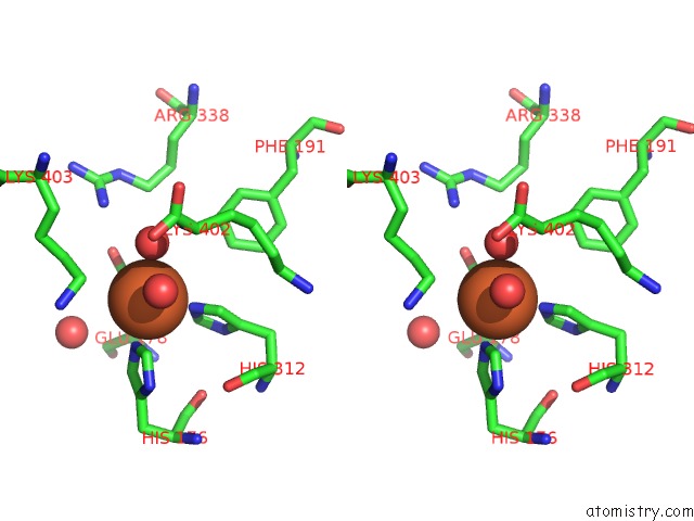
Stereo pair view
Mono view
Stereo pair view
A full contact list of Iron with other atoms in the Fe binding
site number 1 of Crystal Structure of the Complex Fe(II)/Alpha-Ketoglutarate Dependent Dioxygenase KDO5 with Fe(II)/Lysine within 5.0Å range:
|
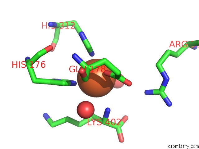
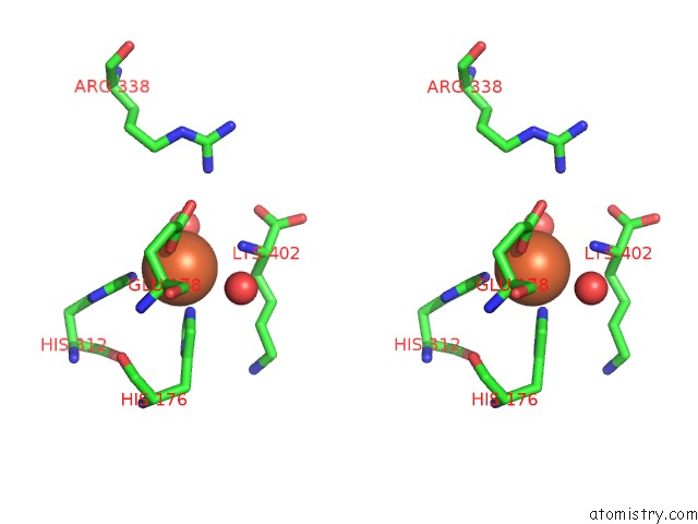
Iron binding site 2 out of 4 in 6exf
Go back to
Iron binding site 2 out
of 4 in the Crystal Structure of the Complex Fe(II)/Alpha-Ketoglutarate Dependent Dioxygenase KDO5 with Fe(II)/Lysine
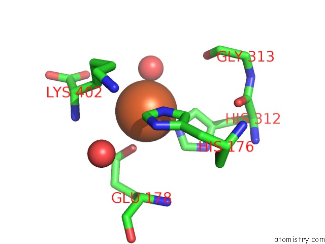
Mono view
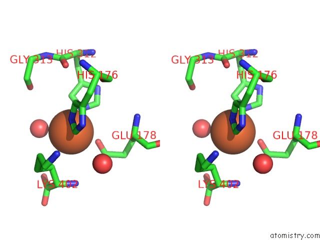
Stereo pair view
Mono view
Stereo pair view
A full contact list of Iron with other atoms in the Fe binding
site number 2 of Crystal Structure of the Complex Fe(II)/Alpha-Ketoglutarate Dependent Dioxygenase KDO5 with Fe(II)/Lysine within 5.0Å range:
|
Iron binding site 3 out of 4 in 6exf
Go back to
Iron binding site 3 out
of 4 in the Crystal Structure of the Complex Fe(II)/Alpha-Ketoglutarate Dependent Dioxygenase KDO5 with Fe(II)/Lysine
Mono view
Stereo pair view
Mono view
Stereo pair view
A full contact list of Iron with other atoms in the Fe binding
site number 3 of Crystal Structure of the Complex Fe(II)/Alpha-Ketoglutarate Dependent Dioxygenase KDO5 with Fe(II)/Lysine within 5.0Å range:
|
Iron binding site 4 out of 4 in 6exf
Go back to
Iron binding site 4 out
of 4 in the Crystal Structure of the Complex Fe(II)/Alpha-Ketoglutarate Dependent Dioxygenase KDO5 with Fe(II)/Lysine
Mono view
Stereo pair view
Mono view
Stereo pair view
A full contact list of Iron with other atoms in the Fe binding
site number 4 of Crystal Structure of the Complex Fe(II)/Alpha-Ketoglutarate Dependent Dioxygenase KDO5 with Fe(II)/Lysine within 5.0Å range:
|
Reference:
K.Bastard,
T.Isabet,
E.A.Stura,
P.Legrand,
A.Zaparucha.
Structural Studies Based on Two Lysine Dioxygenases with Distinct Regioselectivity Brings Insights Into Enzyme Specificity Within the Clavaminate Synthase-Like Family. Sci Rep V. 8 16587 2018.
ISSN: ESSN 2045-2322
PubMed: 30410048
DOI: 10.1038/S41598-018-34795-9
Page generated: Tue Aug 6 17:35:15 2024
ISSN: ESSN 2045-2322
PubMed: 30410048
DOI: 10.1038/S41598-018-34795-9
Last articles
Zn in 9J0NZn in 9J0O
Zn in 9J0P
Zn in 9FJX
Zn in 9EKB
Zn in 9C0F
Zn in 9CAH
Zn in 9CH0
Zn in 9CH3
Zn in 9CH1