Iron »
PDB 6f0b-6fl2 »
6fkt »
Iron in PDB 6fkt: Crystal Structure of A Dye-Decolorizing Peroxidase R232A Variant From Klebsiella Pneumoniae (Kpdyp)
Protein crystallography data
The structure of Crystal Structure of A Dye-Decolorizing Peroxidase R232A Variant From Klebsiella Pneumoniae (Kpdyp), PDB code: 6fkt
was solved by
V.Pfanzagl,
S.Hofbauer,
G.Mlynek,
with X-Ray Crystallography technique. A brief refinement statistics is given in the table below:
| Resolution Low / High (Å) | 75.22 / 1.86 |
| Space group | P 65 2 2 |
| Cell size a, b, c (Å), α, β, γ (°) | 91.160, 91.160, 247.820, 90.00, 90.00, 120.00 |
| R / Rfree (%) | 16.8 / 20.7 |
Other elements in 6fkt:
The structure of Crystal Structure of A Dye-Decolorizing Peroxidase R232A Variant From Klebsiella Pneumoniae (Kpdyp) also contains other interesting chemical elements:
| Magnesium | (Mg) | 1 atom |
Iron Binding Sites:
The binding sites of Iron atom in the Crystal Structure of A Dye-Decolorizing Peroxidase R232A Variant From Klebsiella Pneumoniae (Kpdyp)
(pdb code 6fkt). This binding sites where shown within
5.0 Angstroms radius around Iron atom.
In total 2 binding sites of Iron where determined in the Crystal Structure of A Dye-Decolorizing Peroxidase R232A Variant From Klebsiella Pneumoniae (Kpdyp), PDB code: 6fkt:
Jump to Iron binding site number: 1; 2;
In total 2 binding sites of Iron where determined in the Crystal Structure of A Dye-Decolorizing Peroxidase R232A Variant From Klebsiella Pneumoniae (Kpdyp), PDB code: 6fkt:
Jump to Iron binding site number: 1; 2;
Iron binding site 1 out of 2 in 6fkt
Go back to
Iron binding site 1 out
of 2 in the Crystal Structure of A Dye-Decolorizing Peroxidase R232A Variant From Klebsiella Pneumoniae (Kpdyp)
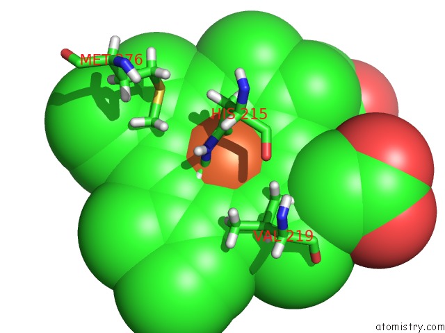
Mono view
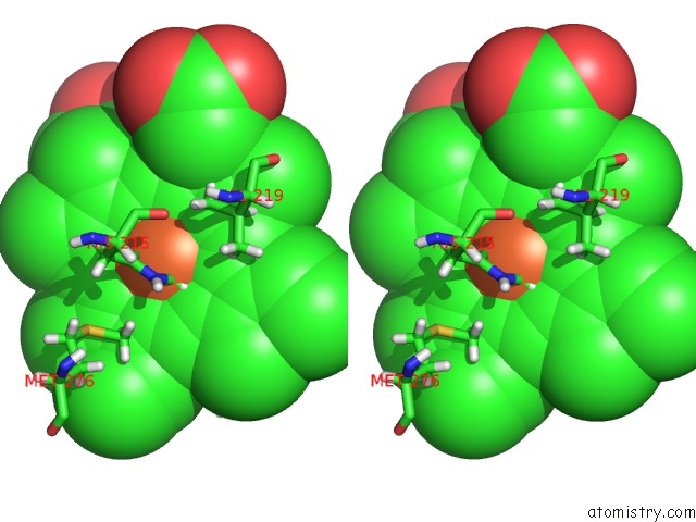
Stereo pair view
Mono view
Stereo pair view
A full contact list of Iron with other atoms in the Fe binding
site number 1 of Crystal Structure of A Dye-Decolorizing Peroxidase R232A Variant From Klebsiella Pneumoniae (Kpdyp) within 5.0Å range:
|
Iron binding site 2 out of 2 in 6fkt
Go back to
Iron binding site 2 out
of 2 in the Crystal Structure of A Dye-Decolorizing Peroxidase R232A Variant From Klebsiella Pneumoniae (Kpdyp)
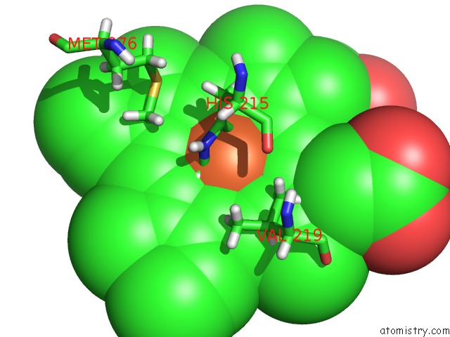
Mono view
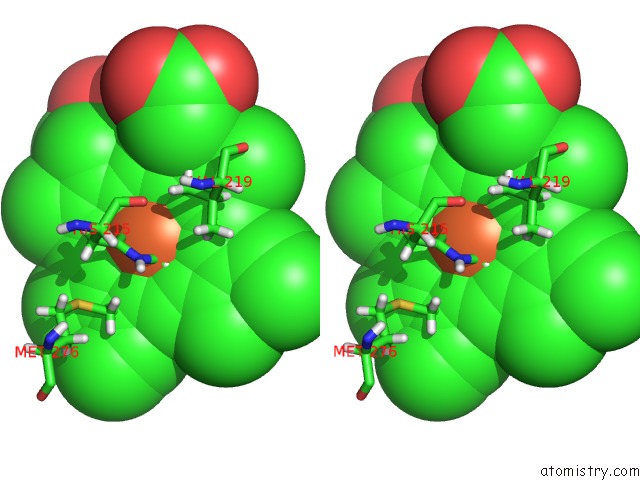
Stereo pair view
Mono view
Stereo pair view
A full contact list of Iron with other atoms in the Fe binding
site number 2 of Crystal Structure of A Dye-Decolorizing Peroxidase R232A Variant From Klebsiella Pneumoniae (Kpdyp) within 5.0Å range:
|
Reference:
V.Pfanzagl,
K.Nys,
M.Bellei,
H.Michlits,
G.Mlynek,
G.Battistuzzi,
K.Djinovic-Carugo,
S.Van Doorslaer,
P.G.Furtmuller,
S.Hofbauer,
C.Obinger.
Roles of Distal Aspartate and Arginine of B-Class Dye-Decolorizing Peroxidase in Heterolytic Hydrogen Peroxide Cleavage. J. Biol. Chem. V. 293 14823 2018.
ISSN: ESSN 1083-351X
PubMed: 30072383
DOI: 10.1074/JBC.RA118.004773
Page generated: Tue Aug 6 18:13:20 2024
ISSN: ESSN 1083-351X
PubMed: 30072383
DOI: 10.1074/JBC.RA118.004773
Last articles
Zn in 9MJ5Zn in 9HNW
Zn in 9G0L
Zn in 9FNE
Zn in 9DZN
Zn in 9E0I
Zn in 9D32
Zn in 9DAK
Zn in 8ZXC
Zn in 8ZUF