Iron »
PDB 6fmo-6g5t »
6fwk »
Iron in PDB 6fwk: The Crystal Structure of POL2CORE-M644G in Complex with Dna and An Incoming Nucleotide
Enzymatic activity of The Crystal Structure of POL2CORE-M644G in Complex with Dna and An Incoming Nucleotide
All present enzymatic activity of The Crystal Structure of POL2CORE-M644G in Complex with Dna and An Incoming Nucleotide:
2.7.7.7;
2.7.7.7;
Protein crystallography data
The structure of The Crystal Structure of POL2CORE-M644G in Complex with Dna and An Incoming Nucleotide, PDB code: 6fwk
was solved by
V.Parkash,
E.Johansson,
with X-Ray Crystallography technique. A brief refinement statistics is given in the table below:
| Resolution Low / High (Å) | 19.99 / 2.50 |
| Space group | P 1 2 1 |
| Cell size a, b, c (Å), α, β, γ (°) | 153.961, 70.342, 158.965, 90.00, 112.82, 90.00 |
| R / Rfree (%) | 22.3 / 26.3 |
Other elements in 6fwk:
The structure of The Crystal Structure of POL2CORE-M644G in Complex with Dna and An Incoming Nucleotide also contains other interesting chemical elements:
| Calcium | (Ca) | 5 atoms |
Iron Binding Sites:
The binding sites of Iron atom in the The Crystal Structure of POL2CORE-M644G in Complex with Dna and An Incoming Nucleotide
(pdb code 6fwk). This binding sites where shown within
5.0 Angstroms radius around Iron atom.
In total 2 binding sites of Iron where determined in the The Crystal Structure of POL2CORE-M644G in Complex with Dna and An Incoming Nucleotide, PDB code: 6fwk:
Jump to Iron binding site number: 1; 2;
In total 2 binding sites of Iron where determined in the The Crystal Structure of POL2CORE-M644G in Complex with Dna and An Incoming Nucleotide, PDB code: 6fwk:
Jump to Iron binding site number: 1; 2;
Iron binding site 1 out of 2 in 6fwk
Go back to
Iron binding site 1 out
of 2 in the The Crystal Structure of POL2CORE-M644G in Complex with Dna and An Incoming Nucleotide
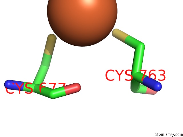
Mono view
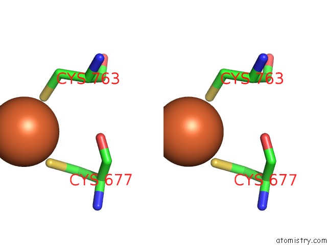
Stereo pair view
Mono view
Stereo pair view
A full contact list of Iron with other atoms in the Fe binding
site number 1 of The Crystal Structure of POL2CORE-M644G in Complex with Dna and An Incoming Nucleotide within 5.0Å range:
|
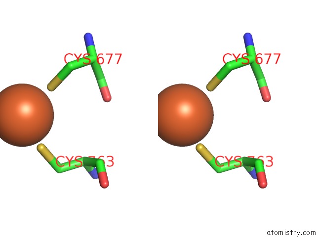
Iron binding site 2 out of 2 in 6fwk
Go back to
Iron binding site 2 out
of 2 in the The Crystal Structure of POL2CORE-M644G in Complex with Dna and An Incoming Nucleotide
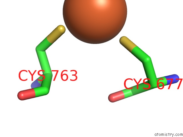
Mono view
Stereo pair view
Mono view
Stereo pair view
A full contact list of Iron with other atoms in the Fe binding
site number 2 of The Crystal Structure of POL2CORE-M644G in Complex with Dna and An Incoming Nucleotide within 5.0Å range:
|
Reference:
V.Parkash,
Y.Kulkarni,
J.Ter Beek,
P.V.Shcherbakova,
S.C.L.Kamerlin,
E.Johansson.
Structural Consequence of the Most Frequently Recurring Cancer-Associated Substitution in Dna Polymerase Epsilon. Nat Commun V. 10 373 2019.
ISSN: ESSN 2041-1723
PubMed: 30670696
DOI: 10.1038/S41467-018-08114-9
Page generated: Tue Aug 6 18:41:10 2024
ISSN: ESSN 2041-1723
PubMed: 30670696
DOI: 10.1038/S41467-018-08114-9
Last articles
Zn in 9J0NZn in 9J0O
Zn in 9J0P
Zn in 9FJX
Zn in 9EKB
Zn in 9C0F
Zn in 9CAH
Zn in 9CH0
Zn in 9CH3
Zn in 9CH1