Iron »
PDB 6fmo-6g5t »
6fxy »
Iron in PDB 6fxy: Crystal Structure of Full-Length Human Lysyl Hydroxylase LH3 - Cocrystal with FE2+, MN2+, Udp-Gal - Structure From Long-Wavelength S-Sad
Enzymatic activity of Crystal Structure of Full-Length Human Lysyl Hydroxylase LH3 - Cocrystal with FE2+, MN2+, Udp-Gal - Structure From Long-Wavelength S-Sad
All present enzymatic activity of Crystal Structure of Full-Length Human Lysyl Hydroxylase LH3 - Cocrystal with FE2+, MN2+, Udp-Gal - Structure From Long-Wavelength S-Sad:
1.14.11.4;
1.14.11.4;
Protein crystallography data
The structure of Crystal Structure of Full-Length Human Lysyl Hydroxylase LH3 - Cocrystal with FE2+, MN2+, Udp-Gal - Structure From Long-Wavelength S-Sad, PDB code: 6fxy
was solved by
L.Scietti,
A.Chiapparino,
F.De Giorgi,
M.Fumagalli,
L.Khoriauli,
S.Nergadze,
S.Basu,
V.Olieric,
B.Banushi,
E.Giulotto,
P.Gissen,
F.Forneris,
with X-Ray Crystallography technique. A brief refinement statistics is given in the table below:
| Resolution Low / High (Å) | 112.37 / 2.14 |
| Space group | C 2 2 21 |
| Cell size a, b, c (Å), α, β, γ (°) | 97.040, 100.105, 224.743, 90.00, 90.00, 90.00 |
| R / Rfree (%) | 19.4 / 23.1 |
Other elements in 6fxy:
The structure of Crystal Structure of Full-Length Human Lysyl Hydroxylase LH3 - Cocrystal with FE2+, MN2+, Udp-Gal - Structure From Long-Wavelength S-Sad also contains other interesting chemical elements:
| Manganese | (Mn) | 1 atom |
Iron Binding Sites:
The binding sites of Iron atom in the Crystal Structure of Full-Length Human Lysyl Hydroxylase LH3 - Cocrystal with FE2+, MN2+, Udp-Gal - Structure From Long-Wavelength S-Sad
(pdb code 6fxy). This binding sites where shown within
5.0 Angstroms radius around Iron atom.
In total 2 binding sites of Iron where determined in the Crystal Structure of Full-Length Human Lysyl Hydroxylase LH3 - Cocrystal with FE2+, MN2+, Udp-Gal - Structure From Long-Wavelength S-Sad, PDB code: 6fxy:
Jump to Iron binding site number: 1; 2;
In total 2 binding sites of Iron where determined in the Crystal Structure of Full-Length Human Lysyl Hydroxylase LH3 - Cocrystal with FE2+, MN2+, Udp-Gal - Structure From Long-Wavelength S-Sad, PDB code: 6fxy:
Jump to Iron binding site number: 1; 2;
Iron binding site 1 out of 2 in 6fxy
Go back to
Iron binding site 1 out
of 2 in the Crystal Structure of Full-Length Human Lysyl Hydroxylase LH3 - Cocrystal with FE2+, MN2+, Udp-Gal - Structure From Long-Wavelength S-Sad
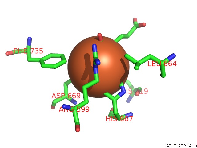
Mono view
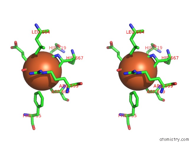
Stereo pair view
Mono view
Stereo pair view
A full contact list of Iron with other atoms in the Fe binding
site number 1 of Crystal Structure of Full-Length Human Lysyl Hydroxylase LH3 - Cocrystal with FE2+, MN2+, Udp-Gal - Structure From Long-Wavelength S-Sad within 5.0Å range:
|
Iron binding site 2 out of 2 in 6fxy
Go back to
Iron binding site 2 out
of 2 in the Crystal Structure of Full-Length Human Lysyl Hydroxylase LH3 - Cocrystal with FE2+, MN2+, Udp-Gal - Structure From Long-Wavelength S-Sad
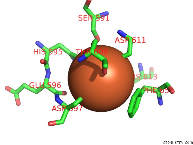
Mono view
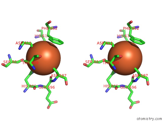
Stereo pair view
Mono view
Stereo pair view
A full contact list of Iron with other atoms in the Fe binding
site number 2 of Crystal Structure of Full-Length Human Lysyl Hydroxylase LH3 - Cocrystal with FE2+, MN2+, Udp-Gal - Structure From Long-Wavelength S-Sad within 5.0Å range:
|
Reference:
L.Scietti,
A.Chiapparino,
F.De Giorgi,
M.Fumagalli,
L.Khoriauli,
S.Nergadze,
S.Basu,
V.Olieric,
L.Cucca,
B.Banushi,
A.Profumo,
E.Giulotto,
P.Gissen,
F.Forneris.
Molecular Architecture of the Multifunctional Collagen Lysyl Hydroxylase and Glycosyltransferase LH3. Nat Commun V. 9 3163 2018.
ISSN: ESSN 2041-1723
PubMed: 30089812
DOI: 10.1038/S41467-018-05631-5
Page generated: Tue Aug 6 18:48:43 2024
ISSN: ESSN 2041-1723
PubMed: 30089812
DOI: 10.1038/S41467-018-05631-5
Last articles
Zn in 9J0NZn in 9J0O
Zn in 9J0P
Zn in 9FJX
Zn in 9EKB
Zn in 9C0F
Zn in 9CAH
Zn in 9CH0
Zn in 9CH3
Zn in 9CH1