Iron »
PDB 6fmo-6g5t »
6g5q »
Iron in PDB 6g5q: The Structure of A Carbohydrate Active P450
Protein crystallography data
The structure of The Structure of A Carbohydrate Active P450, PDB code: 6g5q
was solved by
C.S.Robb,
J.H.Hehemann,
with X-Ray Crystallography technique. A brief refinement statistics is given in the table below:
| Resolution Low / High (Å) | 99.83 / 2.40 |
| Space group | P 21 21 21 |
| Cell size a, b, c (Å), α, β, γ (°) | 63.024, 67.878, 199.653, 90.00, 90.00, 90.00 |
| R / Rfree (%) | 17.2 / 21.7 |
Iron Binding Sites:
The binding sites of Iron atom in the The Structure of A Carbohydrate Active P450
(pdb code 6g5q). This binding sites where shown within
5.0 Angstroms radius around Iron atom.
In total 2 binding sites of Iron where determined in the The Structure of A Carbohydrate Active P450, PDB code: 6g5q:
Jump to Iron binding site number: 1; 2;
In total 2 binding sites of Iron where determined in the The Structure of A Carbohydrate Active P450, PDB code: 6g5q:
Jump to Iron binding site number: 1; 2;
Iron binding site 1 out of 2 in 6g5q
Go back to
Iron binding site 1 out
of 2 in the The Structure of A Carbohydrate Active P450
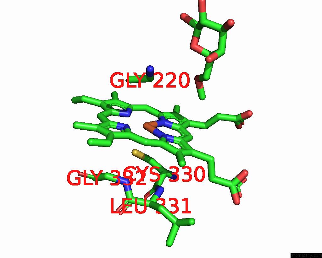
Mono view
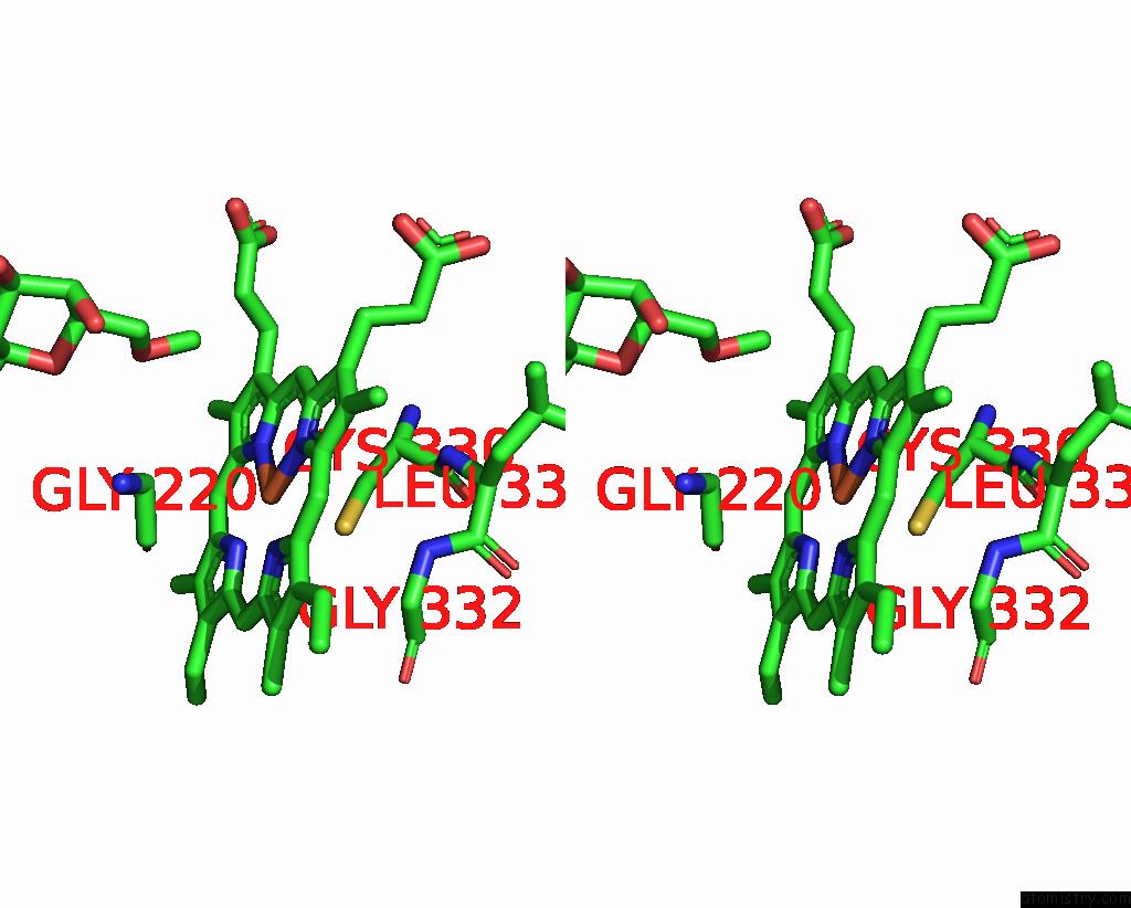
Stereo pair view
Mono view
Stereo pair view
A full contact list of Iron with other atoms in the Fe binding
site number 1 of The Structure of A Carbohydrate Active P450 within 5.0Å range:
|
Iron binding site 2 out of 2 in 6g5q
Go back to
Iron binding site 2 out
of 2 in the The Structure of A Carbohydrate Active P450
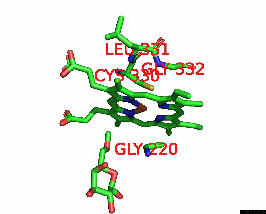
Mono view
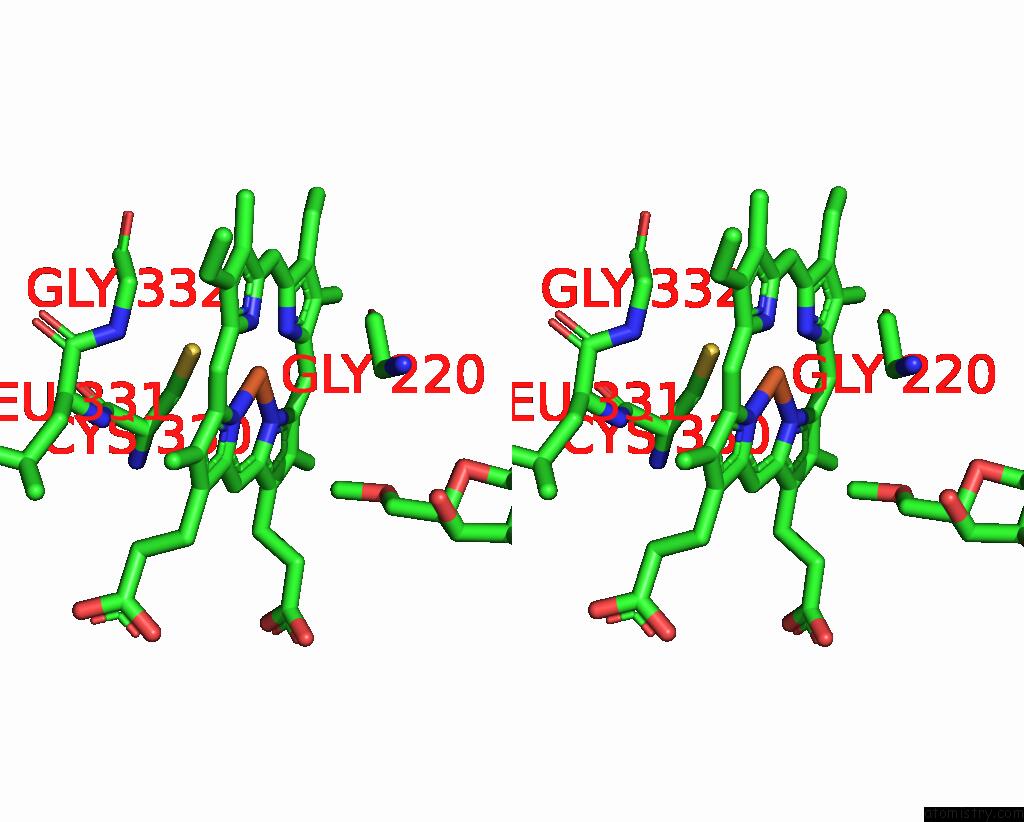
Stereo pair view
Mono view
Stereo pair view
A full contact list of Iron with other atoms in the Fe binding
site number 2 of The Structure of A Carbohydrate Active P450 within 5.0Å range:
|
Reference:
C.S.Robb,
L.Reisky,
U.T.Bornscheuer,
J.H.Hehemann.
Specificity and Mechanism of Carbohydrate Demethylation By Cytochrome P450 Monooxygenases. Biochem. J. V. 475 3875 2018.
ISSN: ESSN 1470-8728
PubMed: 30404923
DOI: 10.1042/BCJ20180762
Page generated: Tue Aug 6 18:55:08 2024
ISSN: ESSN 1470-8728
PubMed: 30404923
DOI: 10.1042/BCJ20180762
Last articles
Zn in 9J0NZn in 9J0O
Zn in 9J0P
Zn in 9FJX
Zn in 9EKB
Zn in 9C0F
Zn in 9CAH
Zn in 9CH0
Zn in 9CH3
Zn in 9CH1