Iron »
PDB 6g71-6gl6 »
6gam »
Iron in PDB 6gam: Structure of E14Q Variant of E. Coli Hydrogenase-2 (As-Isolated Enzyme)
Enzymatic activity of Structure of E14Q Variant of E. Coli Hydrogenase-2 (As-Isolated Enzyme)
All present enzymatic activity of Structure of E14Q Variant of E. Coli Hydrogenase-2 (As-Isolated Enzyme):
1.12.99.6;
1.12.99.6;
Protein crystallography data
The structure of Structure of E14Q Variant of E. Coli Hydrogenase-2 (As-Isolated Enzyme), PDB code: 6gam
was solved by
S.B.Carr,
F.A.Armstrong,
R.M.Evans,
with X-Ray Crystallography technique. A brief refinement statistics is given in the table below:
| Resolution Low / High (Å) | 86.32 / 1.40 |
| Space group | P 21 21 21 |
| Cell size a, b, c (Å), α, β, γ (°) | 99.477, 100.471, 168.656, 90.00, 90.00, 90.00 |
| R / Rfree (%) | 15 / 17 |
Other elements in 6gam:
The structure of Structure of E14Q Variant of E. Coli Hydrogenase-2 (As-Isolated Enzyme) also contains other interesting chemical elements:
| Nickel | (Ni) | 2 atoms |
| Magnesium | (Mg) | 2 atoms |
Iron Binding Sites:
Pages:
>>> Page 1 <<< Page 2, Binding sites: 11 - 20; Page 3, Binding sites: 21 - 24;Binding sites:
The binding sites of Iron atom in the Structure of E14Q Variant of E. Coli Hydrogenase-2 (As-Isolated Enzyme) (pdb code 6gam). This binding sites where shown within 5.0 Angstroms radius around Iron atom.In total 24 binding sites of Iron where determined in the Structure of E14Q Variant of E. Coli Hydrogenase-2 (As-Isolated Enzyme), PDB code: 6gam:
Jump to Iron binding site number: 1; 2; 3; 4; 5; 6; 7; 8; 9; 10;
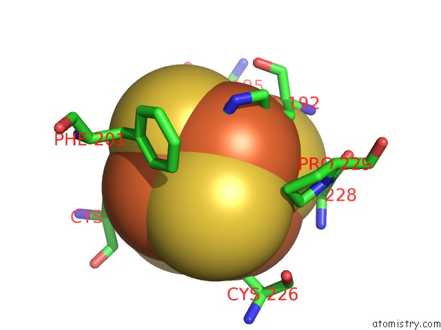
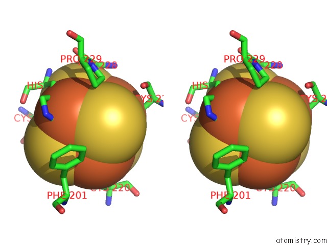
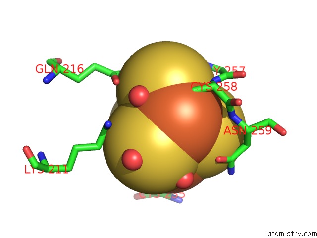
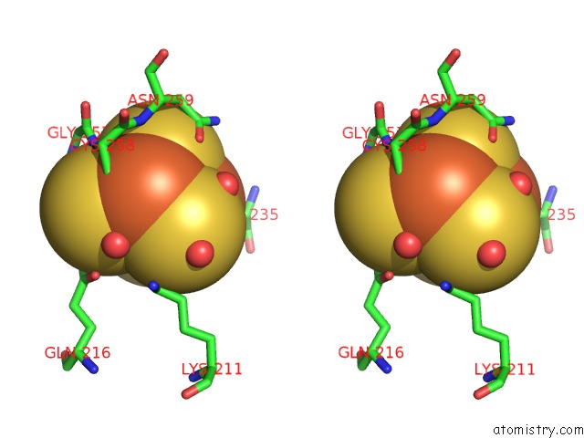
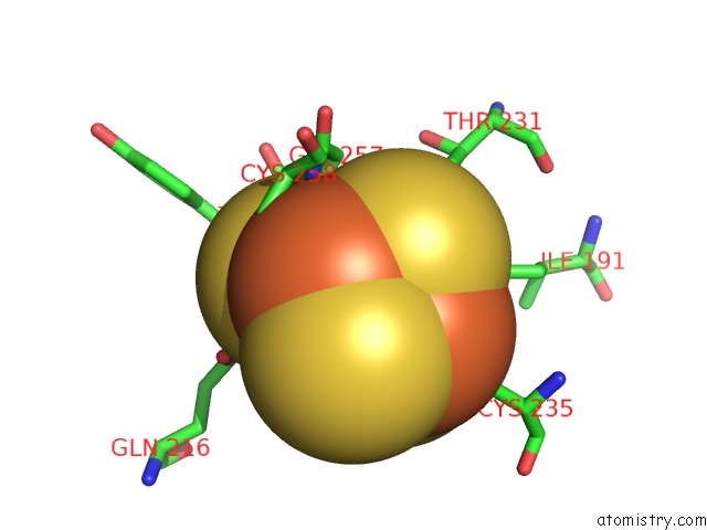
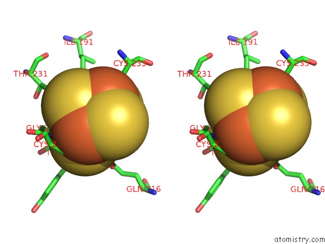
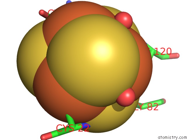
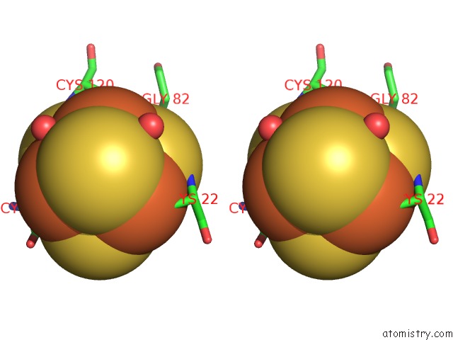
Iron binding site 1 out of 24 in 6gam
Go back to
Iron binding site 1 out
of 24 in the Structure of E14Q Variant of E. Coli Hydrogenase-2 (As-Isolated Enzyme)
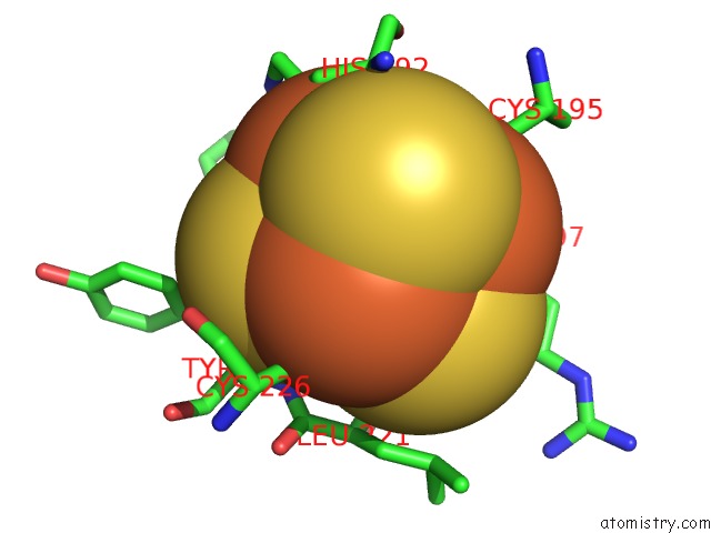
Mono view
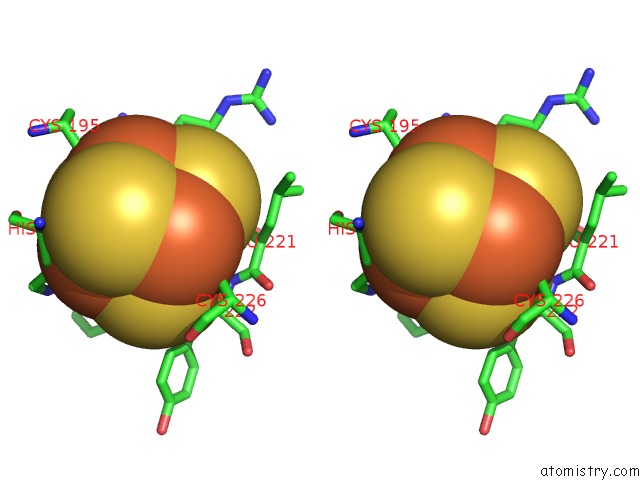
Stereo pair view
Mono view
Stereo pair view
A full contact list of Iron with other atoms in the Fe binding
site number 1 of Structure of E14Q Variant of E. Coli Hydrogenase-2 (As-Isolated Enzyme) within 5.0Å range:
|
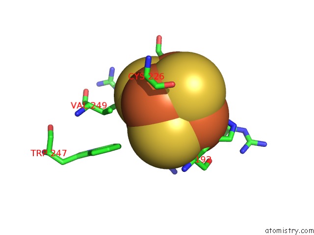
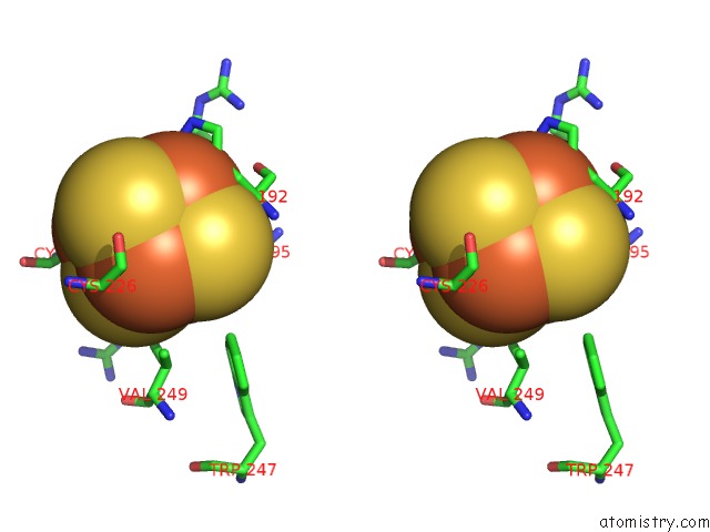
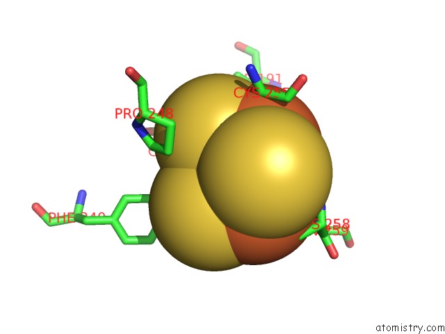
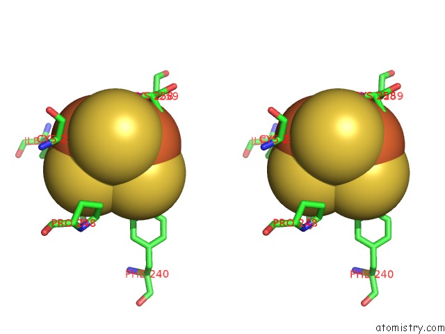
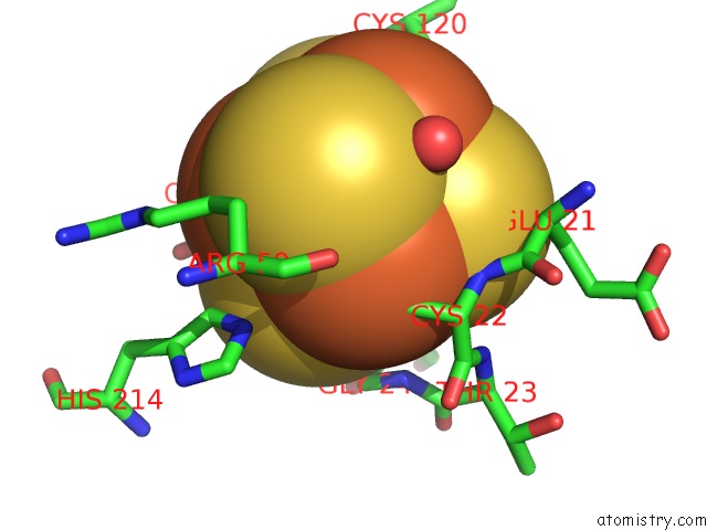
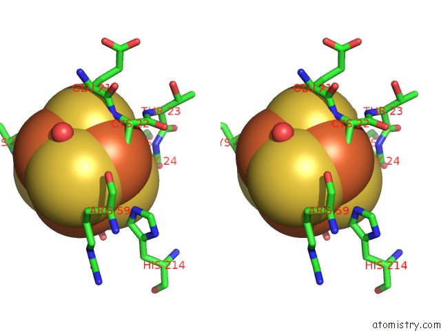
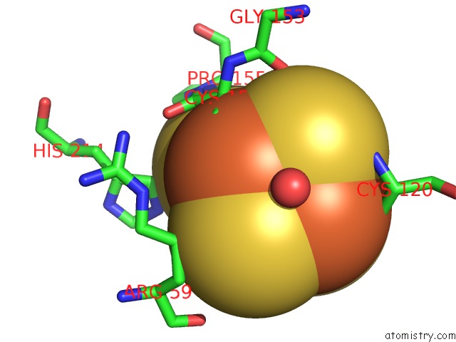
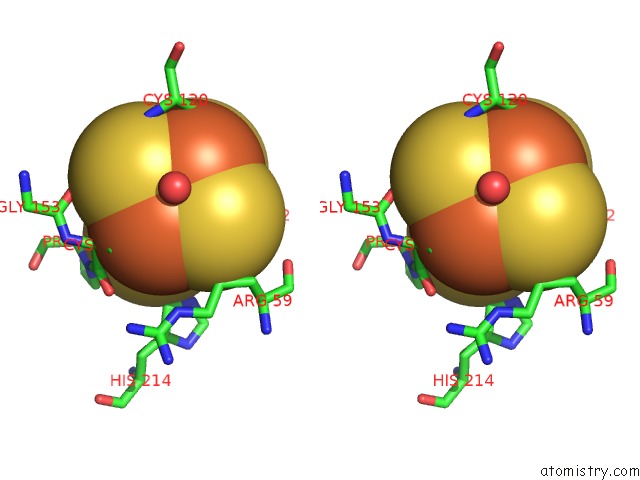
Iron binding site 2 out of 24 in 6gam
Go back to
Iron binding site 2 out
of 24 in the Structure of E14Q Variant of E. Coli Hydrogenase-2 (As-Isolated Enzyme)
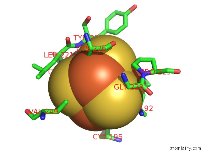
Mono view
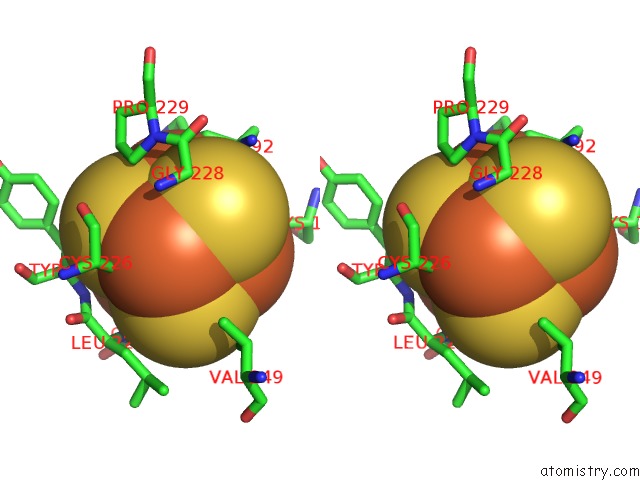
Stereo pair view
Mono view
Stereo pair view
A full contact list of Iron with other atoms in the Fe binding
site number 2 of Structure of E14Q Variant of E. Coli Hydrogenase-2 (As-Isolated Enzyme) within 5.0Å range:
|
Iron binding site 3 out of 24 in 6gam
Go back to
Iron binding site 3 out
of 24 in the Structure of E14Q Variant of E. Coli Hydrogenase-2 (As-Isolated Enzyme)
Mono view
Stereo pair view
Mono view
Stereo pair view
A full contact list of Iron with other atoms in the Fe binding
site number 3 of Structure of E14Q Variant of E. Coli Hydrogenase-2 (As-Isolated Enzyme) within 5.0Å range:
|
Iron binding site 4 out of 24 in 6gam
Go back to
Iron binding site 4 out
of 24 in the Structure of E14Q Variant of E. Coli Hydrogenase-2 (As-Isolated Enzyme)
Mono view
Stereo pair view
Mono view
Stereo pair view
A full contact list of Iron with other atoms in the Fe binding
site number 4 of Structure of E14Q Variant of E. Coli Hydrogenase-2 (As-Isolated Enzyme) within 5.0Å range:
|
Iron binding site 5 out of 24 in 6gam
Go back to
Iron binding site 5 out
of 24 in the Structure of E14Q Variant of E. Coli Hydrogenase-2 (As-Isolated Enzyme)
Mono view
Stereo pair view
Mono view
Stereo pair view
A full contact list of Iron with other atoms in the Fe binding
site number 5 of Structure of E14Q Variant of E. Coli Hydrogenase-2 (As-Isolated Enzyme) within 5.0Å range:
|
Iron binding site 6 out of 24 in 6gam
Go back to
Iron binding site 6 out
of 24 in the Structure of E14Q Variant of E. Coli Hydrogenase-2 (As-Isolated Enzyme)
Mono view
Stereo pair view
Mono view
Stereo pair view
A full contact list of Iron with other atoms in the Fe binding
site number 6 of Structure of E14Q Variant of E. Coli Hydrogenase-2 (As-Isolated Enzyme) within 5.0Å range:
|
Iron binding site 7 out of 24 in 6gam
Go back to
Iron binding site 7 out
of 24 in the Structure of E14Q Variant of E. Coli Hydrogenase-2 (As-Isolated Enzyme)
Mono view
Stereo pair view
Mono view
Stereo pair view
A full contact list of Iron with other atoms in the Fe binding
site number 7 of Structure of E14Q Variant of E. Coli Hydrogenase-2 (As-Isolated Enzyme) within 5.0Å range:
|
Iron binding site 8 out of 24 in 6gam
Go back to
Iron binding site 8 out
of 24 in the Structure of E14Q Variant of E. Coli Hydrogenase-2 (As-Isolated Enzyme)
Mono view
Stereo pair view
Mono view
Stereo pair view
A full contact list of Iron with other atoms in the Fe binding
site number 8 of Structure of E14Q Variant of E. Coli Hydrogenase-2 (As-Isolated Enzyme) within 5.0Å range:
|
Iron binding site 9 out of 24 in 6gam
Go back to
Iron binding site 9 out
of 24 in the Structure of E14Q Variant of E. Coli Hydrogenase-2 (As-Isolated Enzyme)
Mono view
Stereo pair view
Mono view
Stereo pair view
A full contact list of Iron with other atoms in the Fe binding
site number 9 of Structure of E14Q Variant of E. Coli Hydrogenase-2 (As-Isolated Enzyme) within 5.0Å range:
|
Iron binding site 10 out of 24 in 6gam
Go back to
Iron binding site 10 out
of 24 in the Structure of E14Q Variant of E. Coli Hydrogenase-2 (As-Isolated Enzyme)
Mono view
Stereo pair view
Mono view
Stereo pair view
A full contact list of Iron with other atoms in the Fe binding
site number 10 of Structure of E14Q Variant of E. Coli Hydrogenase-2 (As-Isolated Enzyme) within 5.0Å range:
|
Reference:
R.M.Evans,
P.A.Ash,
S.E.Beaton,
E.J.Brooke,
K.A.Vincent,
S.B.Carr,
F.A.Armstrong.
Mechanistic Exploitation of A Self-Repairing, Blocked Proton Transfer Pathway in An O2-Tolerant [Nife]-Hydrogenase. J. Am. Chem. Soc. V. 140 10208 2018.
ISSN: ESSN 1520-5126
PubMed: 30070475
DOI: 10.1021/JACS.8B04798
Page generated: Tue Aug 6 19:13:41 2024
ISSN: ESSN 1520-5126
PubMed: 30070475
DOI: 10.1021/JACS.8B04798
Last articles
F in 7O73F in 7O72
F in 7O70
F in 7O6I
F in 7O4J
F in 7O4I
F in 7O4L
F in 7O4K
F in 7O2V
F in 7NZX