Iron »
PDB 6g71-6gl6 »
6ggu »
Iron in PDB 6ggu: Crystal Structure of Native Fe-Hydrogenase From Methanothermobacter Marburgensis
Enzymatic activity of Crystal Structure of Native Fe-Hydrogenase From Methanothermobacter Marburgensis
All present enzymatic activity of Crystal Structure of Native Fe-Hydrogenase From Methanothermobacter Marburgensis:
1.12.98.2;
1.12.98.2;
Protein crystallography data
The structure of Crystal Structure of Native Fe-Hydrogenase From Methanothermobacter Marburgensis, PDB code: 6ggu
was solved by
T.Wagner,
G.Huang,
U.Ermler,
S.Shima,
with X-Ray Crystallography technique. A brief refinement statistics is given in the table below:
| Resolution Low / High (Å) | 25.84 / 2.60 |
| Space group | P 63 2 2 |
| Cell size a, b, c (Å), α, β, γ (°) | 144.060, 144.060, 95.060, 90.00, 90.00, 120.00 |
| R / Rfree (%) | 18.8 / 23.5 |
Iron Binding Sites:
The binding sites of Iron atom in the Crystal Structure of Native Fe-Hydrogenase From Methanothermobacter Marburgensis
(pdb code 6ggu). This binding sites where shown within
5.0 Angstroms radius around Iron atom.
In total only one binding site of Iron was determined in the Crystal Structure of Native Fe-Hydrogenase From Methanothermobacter Marburgensis, PDB code: 6ggu:
In total only one binding site of Iron was determined in the Crystal Structure of Native Fe-Hydrogenase From Methanothermobacter Marburgensis, PDB code: 6ggu:
Iron binding site 1 out of 1 in 6ggu
Go back to
Iron binding site 1 out
of 1 in the Crystal Structure of Native Fe-Hydrogenase From Methanothermobacter Marburgensis
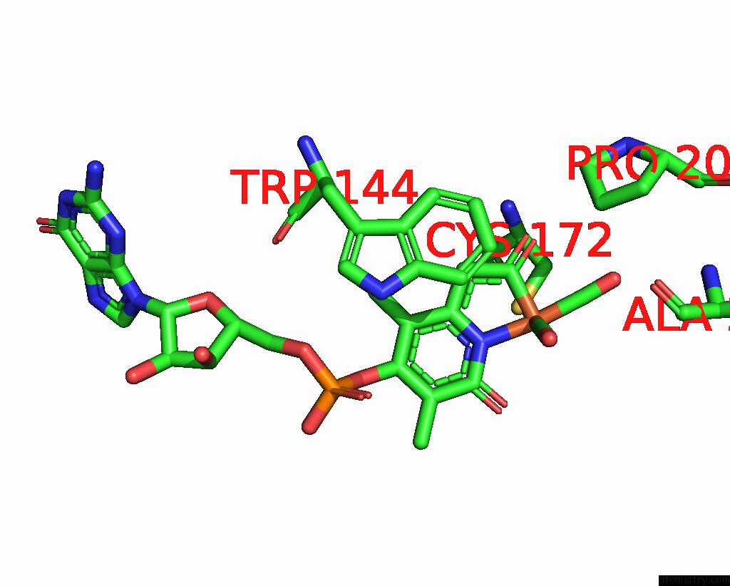
Mono view
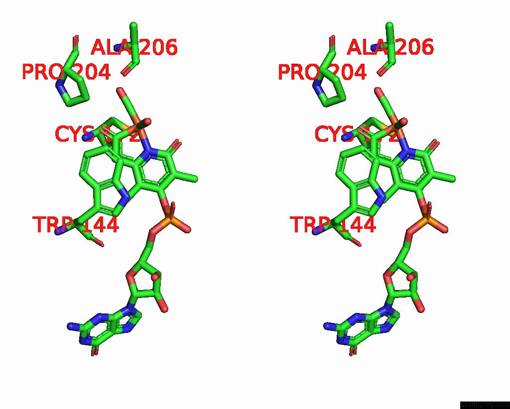
Stereo pair view
Mono view
Stereo pair view
A full contact list of Iron with other atoms in the Fe binding
site number 1 of Crystal Structure of Native Fe-Hydrogenase From Methanothermobacter Marburgensis within 5.0Å range:
|
Reference:
T.Wagner,
G.Huang,
U.Ermler,
S.Shima.
Self-Protection of the Catalytic Iron Center of A Methanogenic [Fe]-Hydrogenase Via A Dynamic Dimer-to-Hexamer Transformation To Be Published.
Page generated: Tue Aug 6 19:30:53 2024
Last articles
Zn in 9JYWZn in 9IR4
Zn in 9IR3
Zn in 9GMX
Zn in 9GMW
Zn in 9JEJ
Zn in 9ERF
Zn in 9ERE
Zn in 9EGV
Zn in 9EGW