Iron »
PDB 6gly-6haw »
6hae »
Iron in PDB 6hae: Crystal Structure of [Fe]-Hydrogenase (Hmd) From Methanococcus Aeolicus in Complex with Fegp Cofactor and Methenyl- Tetrahydromethanopterin (Close Form B)
Enzymatic activity of Crystal Structure of [Fe]-Hydrogenase (Hmd) From Methanococcus Aeolicus in Complex with Fegp Cofactor and Methenyl- Tetrahydromethanopterin (Close Form B)
All present enzymatic activity of Crystal Structure of [Fe]-Hydrogenase (Hmd) From Methanococcus Aeolicus in Complex with Fegp Cofactor and Methenyl- Tetrahydromethanopterin (Close Form B):
1.12.98.2;
1.12.98.2;
Protein crystallography data
The structure of Crystal Structure of [Fe]-Hydrogenase (Hmd) From Methanococcus Aeolicus in Complex with Fegp Cofactor and Methenyl- Tetrahydromethanopterin (Close Form B), PDB code: 6hae
was solved by
G.Huang,
T.Wagner,
M.D.Wodrich,
K.Ataka,
E.Bill,
U.Ermler,
X.Hu,
S.Shima,
with X-Ray Crystallography technique. A brief refinement statistics is given in the table below:
| Resolution Low / High (Å) | 44.56 / 1.85 |
| Space group | P 21 21 2 |
| Cell size a, b, c (Å), α, β, γ (°) | 80.015, 156.480, 53.654, 90.00, 90.00, 90.00 |
| R / Rfree (%) | 16.1 / 19.2 |
Other elements in 6hae:
The structure of Crystal Structure of [Fe]-Hydrogenase (Hmd) From Methanococcus Aeolicus in Complex with Fegp Cofactor and Methenyl- Tetrahydromethanopterin (Close Form B) also contains other interesting chemical elements:
| Potassium | (K) | 4 atoms |
| Chlorine | (Cl) | 2 atoms |
| Sodium | (Na) | 2 atoms |
Iron Binding Sites:
The binding sites of Iron atom in the Crystal Structure of [Fe]-Hydrogenase (Hmd) From Methanococcus Aeolicus in Complex with Fegp Cofactor and Methenyl- Tetrahydromethanopterin (Close Form B)
(pdb code 6hae). This binding sites where shown within
5.0 Angstroms radius around Iron atom.
In total 2 binding sites of Iron where determined in the Crystal Structure of [Fe]-Hydrogenase (Hmd) From Methanococcus Aeolicus in Complex with Fegp Cofactor and Methenyl- Tetrahydromethanopterin (Close Form B), PDB code: 6hae:
Jump to Iron binding site number: 1; 2;
In total 2 binding sites of Iron where determined in the Crystal Structure of [Fe]-Hydrogenase (Hmd) From Methanococcus Aeolicus in Complex with Fegp Cofactor and Methenyl- Tetrahydromethanopterin (Close Form B), PDB code: 6hae:
Jump to Iron binding site number: 1; 2;
Iron binding site 1 out of 2 in 6hae
Go back to
Iron binding site 1 out
of 2 in the Crystal Structure of [Fe]-Hydrogenase (Hmd) From Methanococcus Aeolicus in Complex with Fegp Cofactor and Methenyl- Tetrahydromethanopterin (Close Form B)
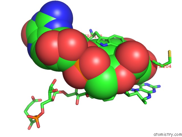
Mono view
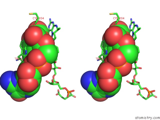
Stereo pair view
Mono view
Stereo pair view
A full contact list of Iron with other atoms in the Fe binding
site number 1 of Crystal Structure of [Fe]-Hydrogenase (Hmd) From Methanococcus Aeolicus in Complex with Fegp Cofactor and Methenyl- Tetrahydromethanopterin (Close Form B) within 5.0Å range:
|
Iron binding site 2 out of 2 in 6hae
Go back to
Iron binding site 2 out
of 2 in the Crystal Structure of [Fe]-Hydrogenase (Hmd) From Methanococcus Aeolicus in Complex with Fegp Cofactor and Methenyl- Tetrahydromethanopterin (Close Form B)
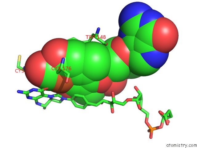
Mono view
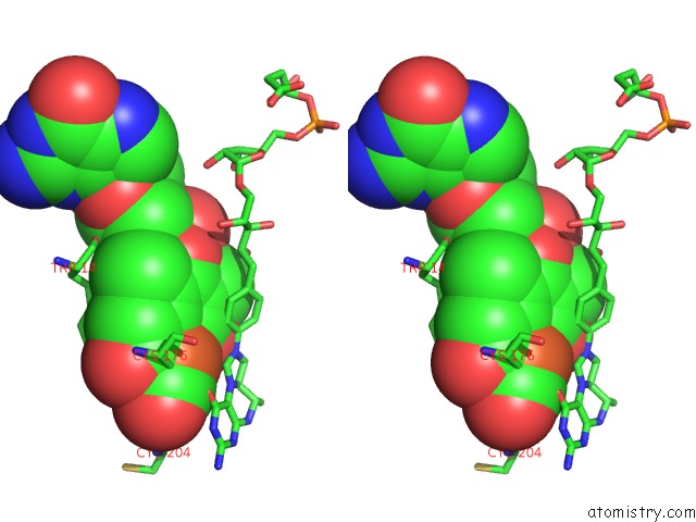
Stereo pair view
Mono view
Stereo pair view
A full contact list of Iron with other atoms in the Fe binding
site number 2 of Crystal Structure of [Fe]-Hydrogenase (Hmd) From Methanococcus Aeolicus in Complex with Fegp Cofactor and Methenyl- Tetrahydromethanopterin (Close Form B) within 5.0Å range:
|
Reference:
G.Huang,
T.Wagner,
M.D.Wodrich,
K.Ataka,
E.Bill,
U.Ermler,
X.Hu,
S.Shima.
The Atomic-Resolution Crystal Structure of Activated [Fe]-Hydrogenase Nat Catal 2019.
ISSN: ESSN 2520-1158
DOI: 10.1038/S41929-019-0289-4
Page generated: Wed Aug 6 07:33:35 2025
ISSN: ESSN 2520-1158
DOI: 10.1038/S41929-019-0289-4
Last articles
Fe in 7MJXFe in 7MJW
Fe in 7MK8
Fe in 7MJZ
Fe in 7MK4
Fe in 7MJY
Fe in 7MI4
Fe in 7MJV
Fe in 7MI5
Fe in 7MID