Iron »
PDB 6hbi-6hsm »
6hmf »
Iron in PDB 6hmf: D-Family Dna Polymerase - DP1 Subunit (3'-5' Proof-Reading Exonuclease) H451 Proof-Reading Deficient Variant
Enzymatic activity of D-Family Dna Polymerase - DP1 Subunit (3'-5' Proof-Reading Exonuclease) H451 Proof-Reading Deficient Variant
All present enzymatic activity of D-Family Dna Polymerase - DP1 Subunit (3'-5' Proof-Reading Exonuclease) H451 Proof-Reading Deficient Variant:
2.7.7.7; 3.1.11.1;
2.7.7.7; 3.1.11.1;
Protein crystallography data
The structure of D-Family Dna Polymerase - DP1 Subunit (3'-5' Proof-Reading Exonuclease) H451 Proof-Reading Deficient Variant, PDB code: 6hmf
was solved by
P.Raia,
M.Delarue,
L.Sauguet,
with X-Ray Crystallography technique. A brief refinement statistics is given in the table below:
| Resolution Low / High (Å) | 47.19 / 2.60 |
| Space group | P 21 21 21 |
| Cell size a, b, c (Å), α, β, γ (°) | 85.750, 91.240, 143.990, 90.00, 90.00, 90.00 |
| R / Rfree (%) | 22.7 / 25.5 |
Other elements in 6hmf:
The structure of D-Family Dna Polymerase - DP1 Subunit (3'-5' Proof-Reading Exonuclease) H451 Proof-Reading Deficient Variant also contains other interesting chemical elements:
| Zinc | (Zn) | 2 atoms |
| Arsenic | (As) | 2 atoms |
| Calcium | (Ca) | 1 atom |
Iron Binding Sites:
The binding sites of Iron atom in the D-Family Dna Polymerase - DP1 Subunit (3'-5' Proof-Reading Exonuclease) H451 Proof-Reading Deficient Variant
(pdb code 6hmf). This binding sites where shown within
5.0 Angstroms radius around Iron atom.
In total 2 binding sites of Iron where determined in the D-Family Dna Polymerase - DP1 Subunit (3'-5' Proof-Reading Exonuclease) H451 Proof-Reading Deficient Variant, PDB code: 6hmf:
Jump to Iron binding site number: 1; 2;
In total 2 binding sites of Iron where determined in the D-Family Dna Polymerase - DP1 Subunit (3'-5' Proof-Reading Exonuclease) H451 Proof-Reading Deficient Variant, PDB code: 6hmf:
Jump to Iron binding site number: 1; 2;
Iron binding site 1 out of 2 in 6hmf
Go back to
Iron binding site 1 out
of 2 in the D-Family Dna Polymerase - DP1 Subunit (3'-5' Proof-Reading Exonuclease) H451 Proof-Reading Deficient Variant
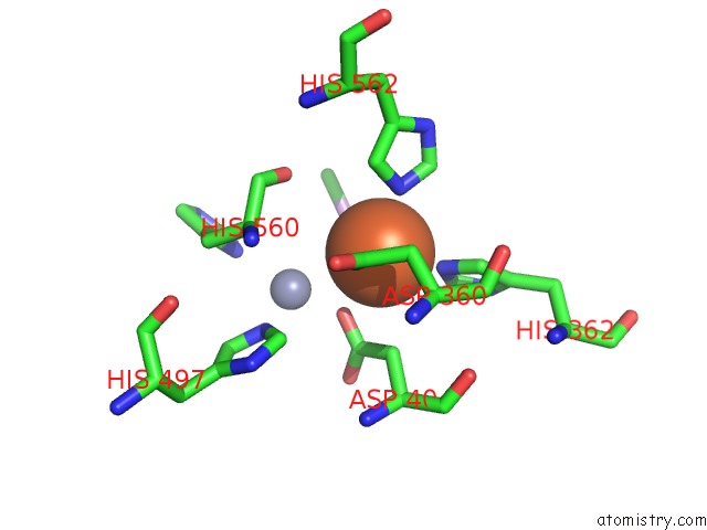
Mono view
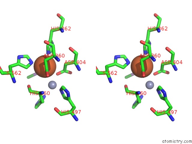
Stereo pair view
Mono view
Stereo pair view
A full contact list of Iron with other atoms in the Fe binding
site number 1 of D-Family Dna Polymerase - DP1 Subunit (3'-5' Proof-Reading Exonuclease) H451 Proof-Reading Deficient Variant within 5.0Å range:
|
Iron binding site 2 out of 2 in 6hmf
Go back to
Iron binding site 2 out
of 2 in the D-Family Dna Polymerase - DP1 Subunit (3'-5' Proof-Reading Exonuclease) H451 Proof-Reading Deficient Variant
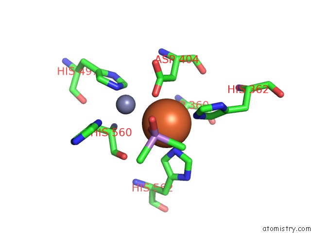
Mono view
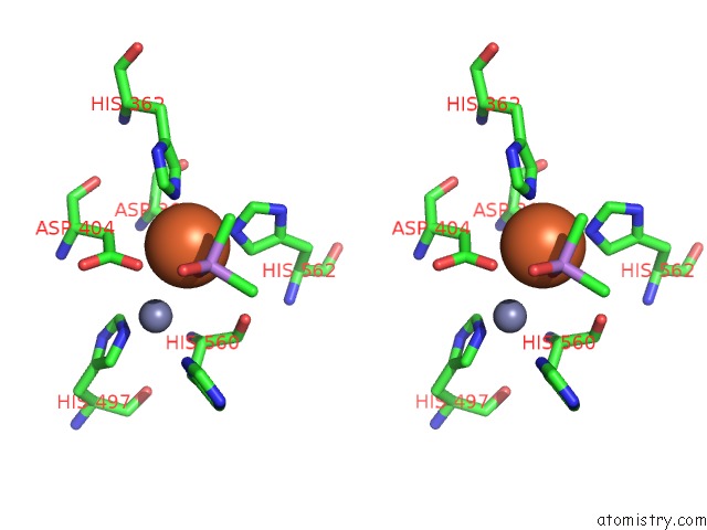
Stereo pair view
Mono view
Stereo pair view
A full contact list of Iron with other atoms in the Fe binding
site number 2 of D-Family Dna Polymerase - DP1 Subunit (3'-5' Proof-Reading Exonuclease) H451 Proof-Reading Deficient Variant within 5.0Å range:
|
Reference:
P.Raia,
M.Carroni,
E.Henry,
G.Pehau-Arnaudet,
S.Brule,
P.Beguin,
G.Henneke,
E.Lindahl,
M.Delarue,
L.Sauguet.
Structure of the DP1-DP2 Pold Complex Bound with Dna and Its Implications For the Evolutionary History of Dna and Rna Polymerases. Plos Biol. V. 17 00122 2019.
ISSN: ESSN 1545-7885
PubMed: 30657780
DOI: 10.1371/JOURNAL.PBIO.3000122
Page generated: Tue Aug 6 21:30:08 2024
ISSN: ESSN 1545-7885
PubMed: 30657780
DOI: 10.1371/JOURNAL.PBIO.3000122
Last articles
Cl in 2XMBCl in 2XMC
Cl in 2XKF
Cl in 2XKQ
Cl in 2XKE
Cl in 2XL7
Cl in 2XKD
Cl in 2XK7
Cl in 2XKC
Cl in 2XK8