Iron »
PDB 6hbi-6hsm »
6hpo »
Iron in PDB 6hpo: Crystallographic Structure of the Catalytic Domain of Human Phenylalanine Hydroxylase (Hpah Cd) in Complex with Iron at 1.6 Angstrom
Enzymatic activity of Crystallographic Structure of the Catalytic Domain of Human Phenylalanine Hydroxylase (Hpah Cd) in Complex with Iron at 1.6 Angstrom
All present enzymatic activity of Crystallographic Structure of the Catalytic Domain of Human Phenylalanine Hydroxylase (Hpah Cd) in Complex with Iron at 1.6 Angstrom:
1.14.16.1;
1.14.16.1;
Protein crystallography data
The structure of Crystallographic Structure of the Catalytic Domain of Human Phenylalanine Hydroxylase (Hpah Cd) in Complex with Iron at 1.6 Angstrom, PDB code: 6hpo
was solved by
M.Alcorlo Pages,
M.Innselset Flydal,
with X-Ray Crystallography technique. A brief refinement statistics is given in the table below:
| Resolution Low / High (Å) | 32.77 / 1.67 |
| Space group | C 2 2 21 |
| Cell size a, b, c (Å), α, β, γ (°) | 65.856, 107.549, 124.016, 90.00, 90.00, 90.00 |
| R / Rfree (%) | 16.1 / 17.7 |
Iron Binding Sites:
The binding sites of Iron atom in the Crystallographic Structure of the Catalytic Domain of Human Phenylalanine Hydroxylase (Hpah Cd) in Complex with Iron at 1.6 Angstrom
(pdb code 6hpo). This binding sites where shown within
5.0 Angstroms radius around Iron atom.
In total only one binding site of Iron was determined in the Crystallographic Structure of the Catalytic Domain of Human Phenylalanine Hydroxylase (Hpah Cd) in Complex with Iron at 1.6 Angstrom, PDB code: 6hpo:
In total only one binding site of Iron was determined in the Crystallographic Structure of the Catalytic Domain of Human Phenylalanine Hydroxylase (Hpah Cd) in Complex with Iron at 1.6 Angstrom, PDB code: 6hpo:
Iron binding site 1 out of 1 in 6hpo
Go back to
Iron binding site 1 out
of 1 in the Crystallographic Structure of the Catalytic Domain of Human Phenylalanine Hydroxylase (Hpah Cd) in Complex with Iron at 1.6 Angstrom
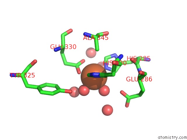
Mono view
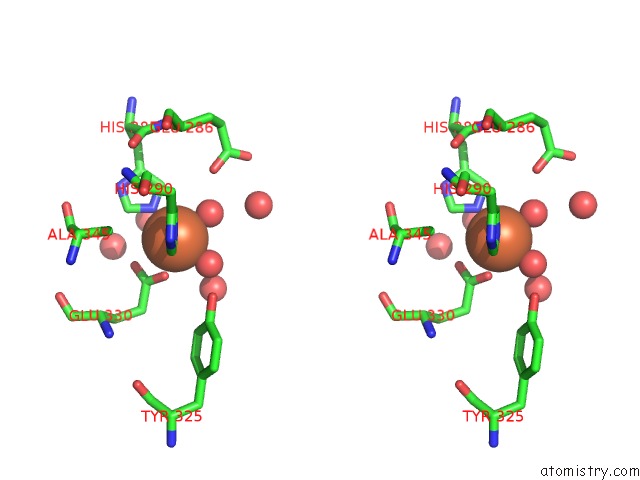
Stereo pair view
Mono view
Stereo pair view
A full contact list of Iron with other atoms in the Fe binding
site number 1 of Crystallographic Structure of the Catalytic Domain of Human Phenylalanine Hydroxylase (Hpah Cd) in Complex with Iron at 1.6 Angstrom within 5.0Å range:
|
Reference:
M.I.Flydal,
M.Alcorlo-Pages,
F.G.Johannessen,
S.Martinez-Caballero,
L.SkjæRven,
R.Fernandez-Leiro,
A.Martinez,
J.A.Hermoso.
Structure of Full-Length Human Phenylalanine Hydroxylase in Complex with Tetrahydrobiopterin. Proc.Natl.Acad.Sci.Usa V. 116 11229 2019.
ISSN: ESSN 1091-6490
PubMed: 31118288
DOI: 10.1073/PNAS.1902639116
Page generated: Tue Aug 6 21:33:02 2024
ISSN: ESSN 1091-6490
PubMed: 31118288
DOI: 10.1073/PNAS.1902639116
Last articles
Zn in 9J0NZn in 9J0O
Zn in 9J0P
Zn in 9FJX
Zn in 9EKB
Zn in 9C0F
Zn in 9CAH
Zn in 9CH0
Zn in 9CH3
Zn in 9CH1