Iron »
PDB 6hbi-6hsm »
6hqd »
Iron in PDB 6hqd: Cytochrome P450-153 From Pseudomonas Sp. 19-Rlim
Protein crystallography data
The structure of Cytochrome P450-153 From Pseudomonas Sp. 19-Rlim, PDB code: 6hqd
was solved by
F.Fiorentini,
A.Mattevi,
with X-Ray Crystallography technique. A brief refinement statistics is given in the table below:
| Resolution Low / High (Å) | 44.00 / 1.80 |
| Space group | P 1 21 1 |
| Cell size a, b, c (Å), α, β, γ (°) | 87.311, 96.642, 87.777, 90.00, 119.18, 90.00 |
| R / Rfree (%) | 17 / 20 |
Iron Binding Sites:
The binding sites of Iron atom in the Cytochrome P450-153 From Pseudomonas Sp. 19-Rlim
(pdb code 6hqd). This binding sites where shown within
5.0 Angstroms radius around Iron atom.
In total 3 binding sites of Iron where determined in the Cytochrome P450-153 From Pseudomonas Sp. 19-Rlim, PDB code: 6hqd:
Jump to Iron binding site number: 1; 2; 3;
In total 3 binding sites of Iron where determined in the Cytochrome P450-153 From Pseudomonas Sp. 19-Rlim, PDB code: 6hqd:
Jump to Iron binding site number: 1; 2; 3;
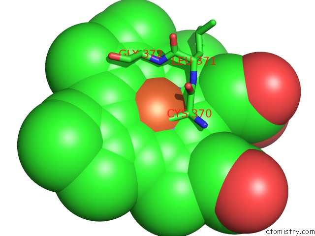
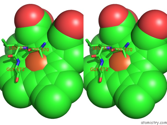
Iron binding site 1 out of 3 in 6hqd
Go back to
Iron binding site 1 out
of 3 in the Cytochrome P450-153 From Pseudomonas Sp. 19-Rlim
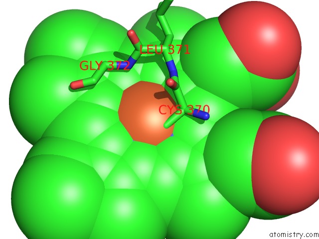
Mono view
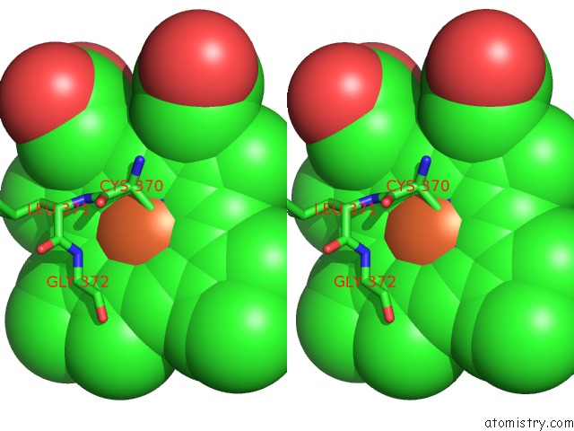
Stereo pair view
Mono view
Stereo pair view
A full contact list of Iron with other atoms in the Fe binding
site number 1 of Cytochrome P450-153 From Pseudomonas Sp. 19-Rlim within 5.0Å range:
|
Iron binding site 2 out of 3 in 6hqd
Go back to
Iron binding site 2 out
of 3 in the Cytochrome P450-153 From Pseudomonas Sp. 19-Rlim
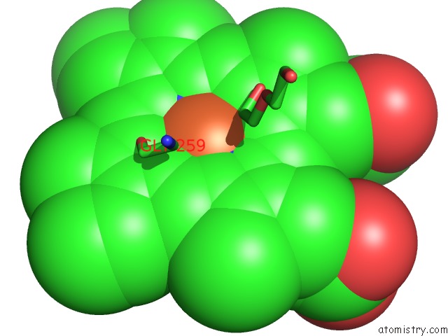
Mono view
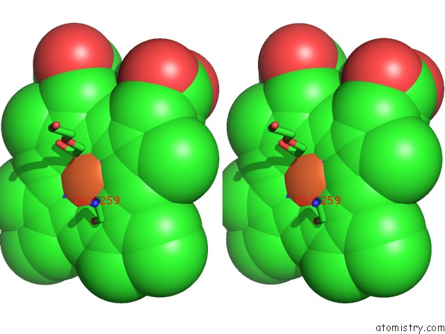
Stereo pair view
Mono view
Stereo pair view
A full contact list of Iron with other atoms in the Fe binding
site number 2 of Cytochrome P450-153 From Pseudomonas Sp. 19-Rlim within 5.0Å range:
|
Iron binding site 3 out of 3 in 6hqd
Go back to
Iron binding site 3 out
of 3 in the Cytochrome P450-153 From Pseudomonas Sp. 19-Rlim
Mono view
Stereo pair view
Mono view
Stereo pair view
A full contact list of Iron with other atoms in the Fe binding
site number 3 of Cytochrome P450-153 From Pseudomonas Sp. 19-Rlim within 5.0Å range:
|
Reference:
F.Fiorentini,
A.M.Hatzl,
S.Schmidt,
S.Savino,
A.Glieder,
A.Mattevi.
The Extreme Structural Plasticity in the CYP153 Subfamily of P450S Directs Development of Designer Hydroxylases. Biochemistry V. 57 6701 2018.
ISSN: ISSN 0006-2960
PubMed: 30398864
DOI: 10.1021/ACS.BIOCHEM.8B01052
Page generated: Tue Aug 6 21:33:50 2024
ISSN: ISSN 0006-2960
PubMed: 30398864
DOI: 10.1021/ACS.BIOCHEM.8B01052
Last articles
Zn in 9J0NZn in 9J0O
Zn in 9J0P
Zn in 9FJX
Zn in 9EKB
Zn in 9C0F
Zn in 9CAH
Zn in 9CH0
Zn in 9CH3
Zn in 9CH1