Iron »
PDB 6hbi-6hsm »
6hqt »
Iron in PDB 6hqt: Crystal Structure of Gcoa F169V Bound to Syringol
Protein crystallography data
The structure of Crystal Structure of Gcoa F169V Bound to Syringol, PDB code: 6hqt
was solved by
S.J.B.Mallinson,
D.J.Hinchen,
M.D.Allen,
C.W.Johnson,
G.T.Beckham,
J.E.Mcgeehan,
with X-Ray Crystallography technique. A brief refinement statistics is given in the table below:
| Resolution Low / High (Å) | 50.50 / 1.85 |
| Space group | P 43 21 2 |
| Cell size a, b, c (Å), α, β, γ (°) | 103.900, 103.900, 115.580, 90.00, 90.00, 90.00 |
| R / Rfree (%) | 15.6 / 17.9 |
Iron Binding Sites:
The binding sites of Iron atom in the Crystal Structure of Gcoa F169V Bound to Syringol
(pdb code 6hqt). This binding sites where shown within
5.0 Angstroms radius around Iron atom.
In total only one binding site of Iron was determined in the Crystal Structure of Gcoa F169V Bound to Syringol, PDB code: 6hqt:
In total only one binding site of Iron was determined in the Crystal Structure of Gcoa F169V Bound to Syringol, PDB code: 6hqt:
Iron binding site 1 out of 1 in 6hqt
Go back to
Iron binding site 1 out
of 1 in the Crystal Structure of Gcoa F169V Bound to Syringol
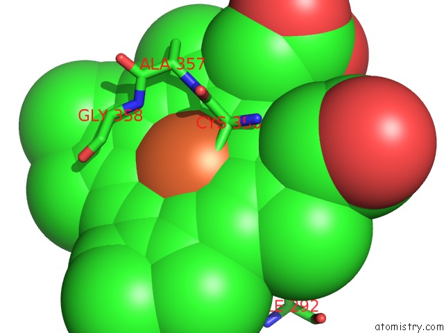
Mono view
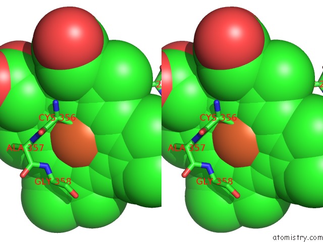
Stereo pair view
Mono view
Stereo pair view
A full contact list of Iron with other atoms in the Fe binding
site number 1 of Crystal Structure of Gcoa F169V Bound to Syringol within 5.0Å range:
|
Reference:
M.M.Machovina,
S.J.B.Mallinson,
B.C.Knott,
A.W.Meyers,
M.Garcia-Borras,
L.Bu,
J.E.Gado,
A.Oliver,
G.P.Schmidt,
D.J.Hinchen,
M.F.Crowley,
C.W.Johnson,
E.L.Neidle,
C.M.Payne,
K.N.Houk,
G.T.Beckham,
J.E.Mcgeehan,
J.L.Dubois.
Enabling Microbial Syringol Conversion Through Structure-Guided Protein Engineering. Proc.Natl.Acad.Sci.Usa V. 116 13970 2019.
ISSN: ESSN 1091-6490
PubMed: 31235604
DOI: 10.1073/PNAS.1820001116
Page generated: Tue Aug 6 21:44:14 2024
ISSN: ESSN 1091-6490
PubMed: 31235604
DOI: 10.1073/PNAS.1820001116
Last articles
Zn in 9J0NZn in 9J0O
Zn in 9J0P
Zn in 9FJX
Zn in 9EKB
Zn in 9C0F
Zn in 9CAH
Zn in 9CH0
Zn in 9CH3
Zn in 9CH1