Iron »
PDB 6htk-6i93 »
6i36 »
Iron in PDB 6i36: Sixty Minutes Iron Loaded Frog M Ferritin
Enzymatic activity of Sixty Minutes Iron Loaded Frog M Ferritin
All present enzymatic activity of Sixty Minutes Iron Loaded Frog M Ferritin:
1.16.3.1;
1.16.3.1;
Protein crystallography data
The structure of Sixty Minutes Iron Loaded Frog M Ferritin, PDB code: 6i36
was solved by
S.Mangani,
F.Di Pisa,
C.Pozzi,
P.Turano,
D.Lalli,
with X-Ray Crystallography technique. A brief refinement statistics is given in the table below:
| Resolution Low / High (Å) | 19.64 / 1.59 |
| Space group | F 4 3 2 |
| Cell size a, b, c (Å), α, β, γ (°) | 184.200, 184.200, 184.200, 90.00, 90.00, 90.00 |
| R / Rfree (%) | 19.2 / 20.5 |
Other elements in 6i36:
The structure of Sixty Minutes Iron Loaded Frog M Ferritin also contains other interesting chemical elements:
| Magnesium | (Mg) | 4 atoms |
| Chlorine | (Cl) | 15 atoms |
Iron Binding Sites:
The binding sites of Iron atom in the Sixty Minutes Iron Loaded Frog M Ferritin
(pdb code 6i36). This binding sites where shown within
5.0 Angstroms radius around Iron atom.
In total 6 binding sites of Iron where determined in the Sixty Minutes Iron Loaded Frog M Ferritin, PDB code: 6i36:
Jump to Iron binding site number: 1; 2; 3; 4; 5; 6;
In total 6 binding sites of Iron where determined in the Sixty Minutes Iron Loaded Frog M Ferritin, PDB code: 6i36:
Jump to Iron binding site number: 1; 2; 3; 4; 5; 6;
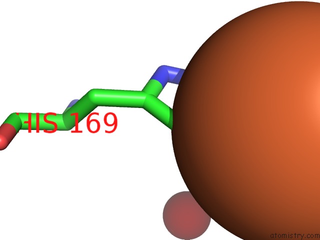
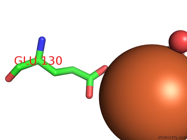
Iron binding site 1 out of 6 in 6i36
Go back to
Iron binding site 1 out
of 6 in the Sixty Minutes Iron Loaded Frog M Ferritin
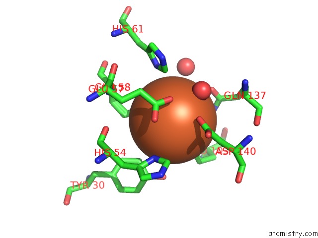
Mono view
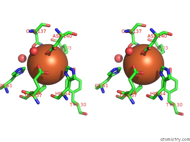
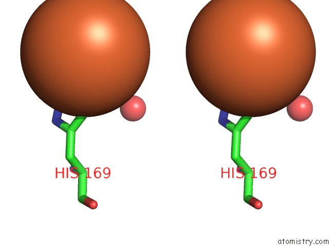
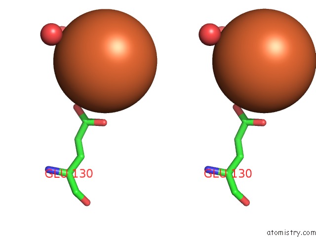
Stereo pair view
Mono view
Stereo pair view
A full contact list of Iron with other atoms in the Fe binding
site number 1 of Sixty Minutes Iron Loaded Frog M Ferritin within 5.0Å range:
|
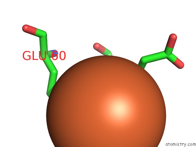
Iron binding site 2 out of 6 in 6i36
Go back to
Iron binding site 2 out
of 6 in the Sixty Minutes Iron Loaded Frog M Ferritin
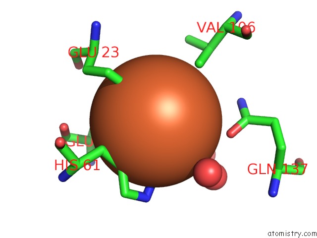
Mono view
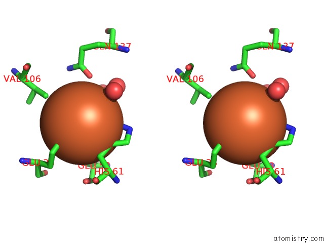
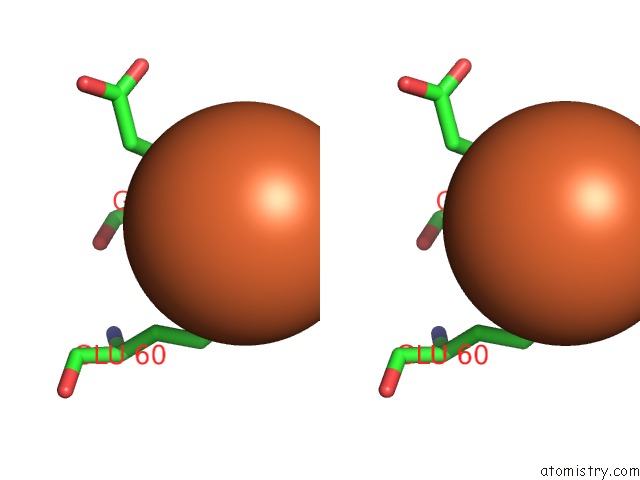
Stereo pair view
Mono view
Stereo pair view
A full contact list of Iron with other atoms in the Fe binding
site number 2 of Sixty Minutes Iron Loaded Frog M Ferritin within 5.0Å range:
|
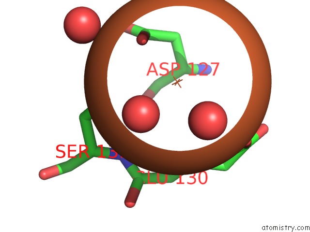
Iron binding site 3 out of 6 in 6i36
Go back to
Iron binding site 3 out
of 6 in the Sixty Minutes Iron Loaded Frog M Ferritin
Mono view
Stereo pair view
Mono view
Stereo pair view
A full contact list of Iron with other atoms in the Fe binding
site number 3 of Sixty Minutes Iron Loaded Frog M Ferritin within 5.0Å range:
|
Iron binding site 4 out of 6 in 6i36
Go back to
Iron binding site 4 out
of 6 in the Sixty Minutes Iron Loaded Frog M Ferritin
Mono view
Stereo pair view
Mono view
Stereo pair view
A full contact list of Iron with other atoms in the Fe binding
site number 4 of Sixty Minutes Iron Loaded Frog M Ferritin within 5.0Å range:
|
Iron binding site 5 out of 6 in 6i36
Go back to
Iron binding site 5 out
of 6 in the Sixty Minutes Iron Loaded Frog M Ferritin
Mono view
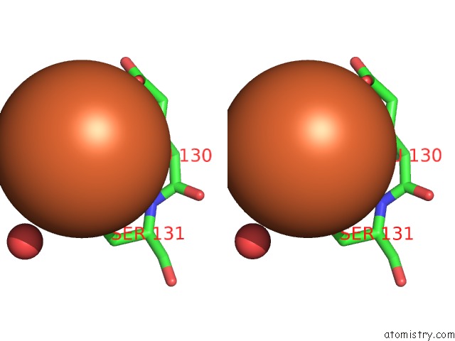
Stereo pair view
Mono view
Stereo pair view
A full contact list of Iron with other atoms in the Fe binding
site number 5 of Sixty Minutes Iron Loaded Frog M Ferritin within 5.0Å range:
|
Iron binding site 6 out of 6 in 6i36
Go back to
Iron binding site 6 out
of 6 in the Sixty Minutes Iron Loaded Frog M Ferritin
Mono view
Stereo pair view
Mono view
Stereo pair view
A full contact list of Iron with other atoms in the Fe binding
site number 6 of Sixty Minutes Iron Loaded Frog M Ferritin within 5.0Å range:
|
Reference:
C.Pozzi,
F.Di Pisa,
D.Lalli,
C.Rosa,
E.Theil,
P.Turano,
S.Mangani.
Time-Lapse Anomalous X-Ray Diffraction Shows How Fe(2+) Substrate Ions Move Through Ferritin Protein Nanocages to Oxidoreductase Sites. Acta Crystallogr.,Sect.D V. 71 941 2015.
ISSN: ISSN 0907-4449
PubMed: 25849404
DOI: 10.1107/S1399004715002333
Page generated: Tue Aug 6 22:03:58 2024
ISSN: ISSN 0907-4449
PubMed: 25849404
DOI: 10.1107/S1399004715002333
Last articles
Zn in 9MJ5Zn in 9HNW
Zn in 9G0L
Zn in 9FNE
Zn in 9DZN
Zn in 9E0I
Zn in 9D32
Zn in 9DAK
Zn in 8ZXC
Zn in 8ZUF