Iron »
PDB 6i95-6j55 »
6iao »
Iron in PDB 6iao: Structure of Cytochrome P450 BM3 M11 Mutant in Complex with Dtt at Resolution 2.16A
Enzymatic activity of Structure of Cytochrome P450 BM3 M11 Mutant in Complex with Dtt at Resolution 2.16A
All present enzymatic activity of Structure of Cytochrome P450 BM3 M11 Mutant in Complex with Dtt at Resolution 2.16A:
1.14.14.1; 1.6.2.4;
1.14.14.1; 1.6.2.4;
Protein crystallography data
The structure of Structure of Cytochrome P450 BM3 M11 Mutant in Complex with Dtt at Resolution 2.16A, PDB code: 6iao
was solved by
O.Mirza,
M.Rafiq,
K.Frydenvang,
with X-Ray Crystallography technique. A brief refinement statistics is given in the table below:
| Resolution Low / High (Å) | 47.18 / 2.16 |
| Space group | C 1 2 1 |
| Cell size a, b, c (Å), α, β, γ (°) | 377.933, 59.937, 95.456, 90.00, 95.74, 90.00 |
| R / Rfree (%) | 15.9 / 20.2 |
Other elements in 6iao:
The structure of Structure of Cytochrome P450 BM3 M11 Mutant in Complex with Dtt at Resolution 2.16A also contains other interesting chemical elements:
| Chlorine | (Cl) | 20 atoms |
Iron Binding Sites:
The binding sites of Iron atom in the Structure of Cytochrome P450 BM3 M11 Mutant in Complex with Dtt at Resolution 2.16A
(pdb code 6iao). This binding sites where shown within
5.0 Angstroms radius around Iron atom.
In total 4 binding sites of Iron where determined in the Structure of Cytochrome P450 BM3 M11 Mutant in Complex with Dtt at Resolution 2.16A, PDB code: 6iao:
Jump to Iron binding site number: 1; 2; 3; 4;
In total 4 binding sites of Iron where determined in the Structure of Cytochrome P450 BM3 M11 Mutant in Complex with Dtt at Resolution 2.16A, PDB code: 6iao:
Jump to Iron binding site number: 1; 2; 3; 4;
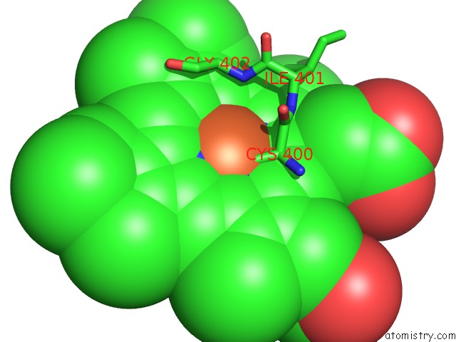
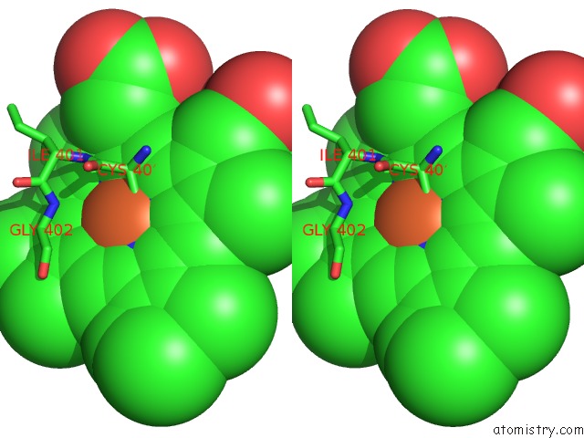
Iron binding site 1 out of 4 in 6iao
Go back to
Iron binding site 1 out
of 4 in the Structure of Cytochrome P450 BM3 M11 Mutant in Complex with Dtt at Resolution 2.16A
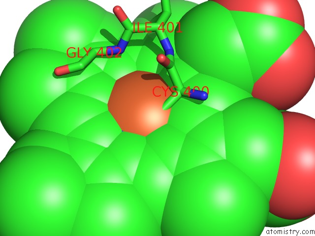
Mono view
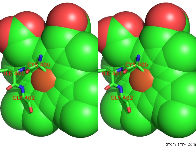
Stereo pair view
Mono view
Stereo pair view
A full contact list of Iron with other atoms in the Fe binding
site number 1 of Structure of Cytochrome P450 BM3 M11 Mutant in Complex with Dtt at Resolution 2.16A within 5.0Å range:
|
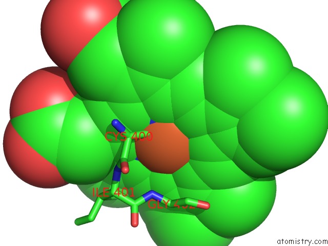
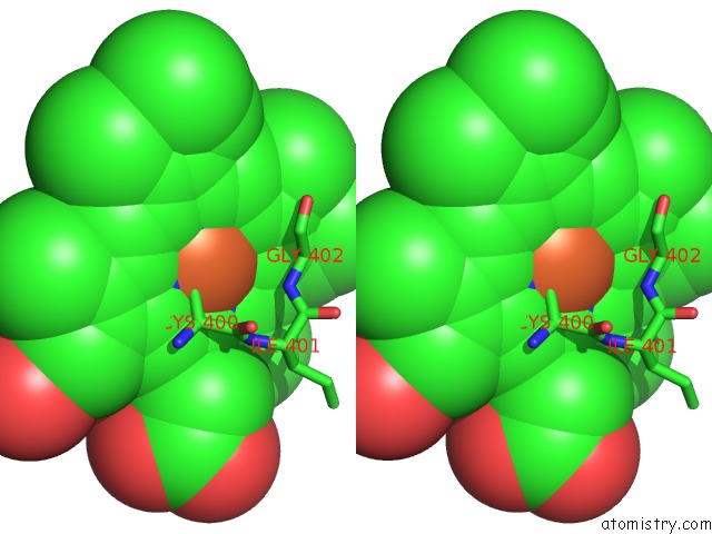
Iron binding site 2 out of 4 in 6iao
Go back to
Iron binding site 2 out
of 4 in the Structure of Cytochrome P450 BM3 M11 Mutant in Complex with Dtt at Resolution 2.16A
Mono view
Stereo pair view
Mono view
Stereo pair view
A full contact list of Iron with other atoms in the Fe binding
site number 2 of Structure of Cytochrome P450 BM3 M11 Mutant in Complex with Dtt at Resolution 2.16A within 5.0Å range:
|
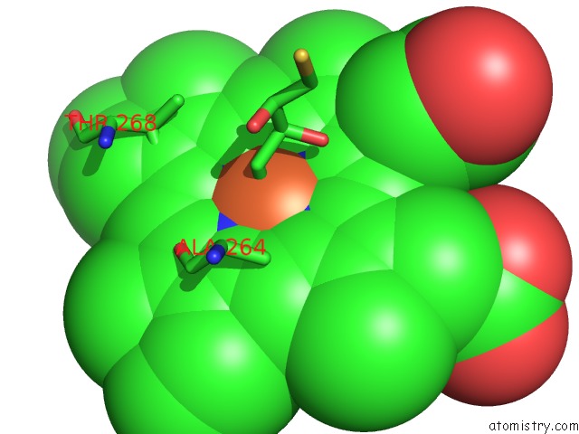
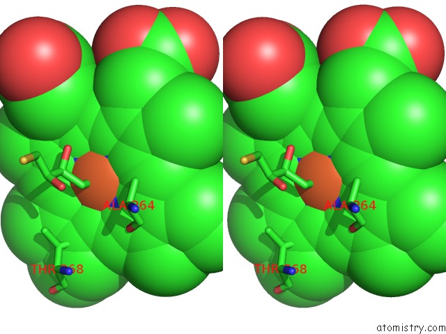
Iron binding site 3 out of 4 in 6iao
Go back to
Iron binding site 3 out
of 4 in the Structure of Cytochrome P450 BM3 M11 Mutant in Complex with Dtt at Resolution 2.16A
Mono view
Stereo pair view
Mono view
Stereo pair view
A full contact list of Iron with other atoms in the Fe binding
site number 3 of Structure of Cytochrome P450 BM3 M11 Mutant in Complex with Dtt at Resolution 2.16A within 5.0Å range:
|
Iron binding site 4 out of 4 in 6iao
Go back to
Iron binding site 4 out
of 4 in the Structure of Cytochrome P450 BM3 M11 Mutant in Complex with Dtt at Resolution 2.16A
Mono view
Stereo pair view
Mono view
Stereo pair view
A full contact list of Iron with other atoms in the Fe binding
site number 4 of Structure of Cytochrome P450 BM3 M11 Mutant in Complex with Dtt at Resolution 2.16A within 5.0Å range:
|
Reference:
K.Frydenvang,
M.C.A.Verkade-Vreeker,
F.Dohmen,
J.N.M.Commandeur,
M.Rafiq,
O.Mirza,
F.S.Jorgensen,
D.P.Geerke.
Structural Analysis of Cytochrome P450 BM3 Mutant M11 in Complex with Dithiothreitol. Plos One V. 14 17292 2019.
ISSN: ESSN 1932-6203
PubMed: 31125381
DOI: 10.1371/JOURNAL.PONE.0217292
Page generated: Wed Aug 6 08:20:01 2025
ISSN: ESSN 1932-6203
PubMed: 31125381
DOI: 10.1371/JOURNAL.PONE.0217292
Last articles
Fe in 6N1FFe in 6N0K
Fe in 6N0J
Fe in 6N03
Fe in 6N02
Fe in 6MYS
Fe in 6MYR
Fe in 6MYQ
Fe in 6MSO
Fe in 6MYP