Iron »
PDB 6jsu-6kbh »
6k5z »
Iron in PDB 6k5z: Structure of Uridylyltransferase
Protein crystallography data
The structure of Structure of Uridylyltransferase, PDB code: 6k5z
was solved by
H.Sakuraba,
T.Ohshida,
K.Yoneda,
T.Ohshima,
with X-Ray Crystallography technique. A brief refinement statistics is given in the table below:
| Resolution Low / High (Å) | 50.00 / 2.33 |
| Space group | P 43 |
| Cell size a, b, c (Å), α, β, γ (°) | 73.194, 73.194, 126.036, 90.00, 90.00, 90.00 |
| R / Rfree (%) | 19.2 / 25.5 |
Other elements in 6k5z:
The structure of Structure of Uridylyltransferase also contains other interesting chemical elements:
| Zinc | (Zn) | 4 atoms |
Iron Binding Sites:
The binding sites of Iron atom in the Structure of Uridylyltransferase
(pdb code 6k5z). This binding sites where shown within
5.0 Angstroms radius around Iron atom.
In total 2 binding sites of Iron where determined in the Structure of Uridylyltransferase, PDB code: 6k5z:
Jump to Iron binding site number: 1; 2;
In total 2 binding sites of Iron where determined in the Structure of Uridylyltransferase, PDB code: 6k5z:
Jump to Iron binding site number: 1; 2;
Iron binding site 1 out of 2 in 6k5z
Go back to
Iron binding site 1 out
of 2 in the Structure of Uridylyltransferase
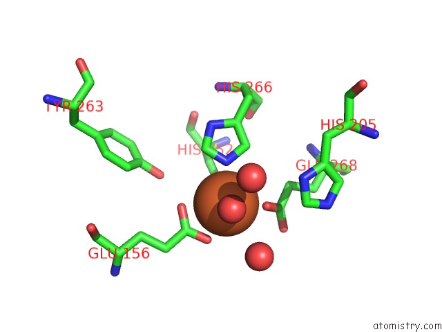
Mono view
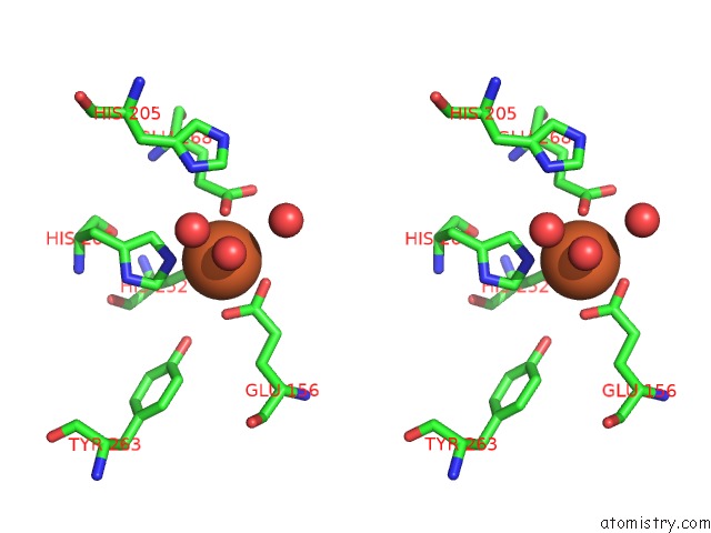
Stereo pair view
Mono view
Stereo pair view
A full contact list of Iron with other atoms in the Fe binding
site number 1 of Structure of Uridylyltransferase within 5.0Å range:
|
Iron binding site 2 out of 2 in 6k5z
Go back to
Iron binding site 2 out
of 2 in the Structure of Uridylyltransferase
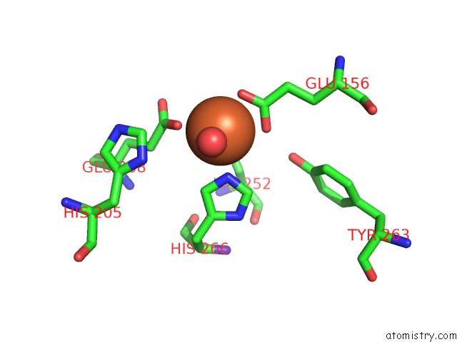
Mono view
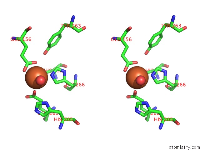
Stereo pair view
Mono view
Stereo pair view
A full contact list of Iron with other atoms in the Fe binding
site number 2 of Structure of Uridylyltransferase within 5.0Å range:
|
Reference:
T.Ohshida,
J.Hayashi,
K.Yoneda,
T.Ohshima,
H.Sakuraba.
Unique Active Site Formation in A Novel Galactose 1-Phosphate Uridylyltransferase From the Hyperthermophilic Archaeon Pyrobaculum Aerophilum. Proteins 2019.
ISSN: ESSN 1097-0134
PubMed: 31693208
DOI: 10.1002/PROT.25848
Page generated: Tue Aug 6 23:46:19 2024
ISSN: ESSN 1097-0134
PubMed: 31693208
DOI: 10.1002/PROT.25848
Last articles
Zn in 9MJ5Zn in 9HNW
Zn in 9G0L
Zn in 9FNE
Zn in 9DZN
Zn in 9E0I
Zn in 9D32
Zn in 9DAK
Zn in 8ZXC
Zn in 8ZUF