Iron »
PDB 6jsu-6kbh »
6kav »
Iron in PDB 6kav: Carbonmonoxy Human Hemoglobin A in the R2 Quaternary Structure at 140 K: Light
Protein crystallography data
The structure of Carbonmonoxy Human Hemoglobin A in the R2 Quaternary Structure at 140 K: Light, PDB code: 6kav
was solved by
N.Shibayama,
S.Y.Park,
M.Ohki,
A.Sato-Tomita,
with X-Ray Crystallography technique. A brief refinement statistics is given in the table below:
| Resolution Low / High (Å) | 19.77 / 1.70 |
| Space group | P 21 21 21 |
| Cell size a, b, c (Å), α, β, γ (°) | 61.146, 96.527, 100.224, 90.00, 90.00, 90.00 |
| R / Rfree (%) | 17.5 / 20.5 |
Iron Binding Sites:
The binding sites of Iron atom in the Carbonmonoxy Human Hemoglobin A in the R2 Quaternary Structure at 140 K: Light
(pdb code 6kav). This binding sites where shown within
5.0 Angstroms radius around Iron atom.
In total 4 binding sites of Iron where determined in the Carbonmonoxy Human Hemoglobin A in the R2 Quaternary Structure at 140 K: Light, PDB code: 6kav:
Jump to Iron binding site number: 1; 2; 3; 4;
In total 4 binding sites of Iron where determined in the Carbonmonoxy Human Hemoglobin A in the R2 Quaternary Structure at 140 K: Light, PDB code: 6kav:
Jump to Iron binding site number: 1; 2; 3; 4;
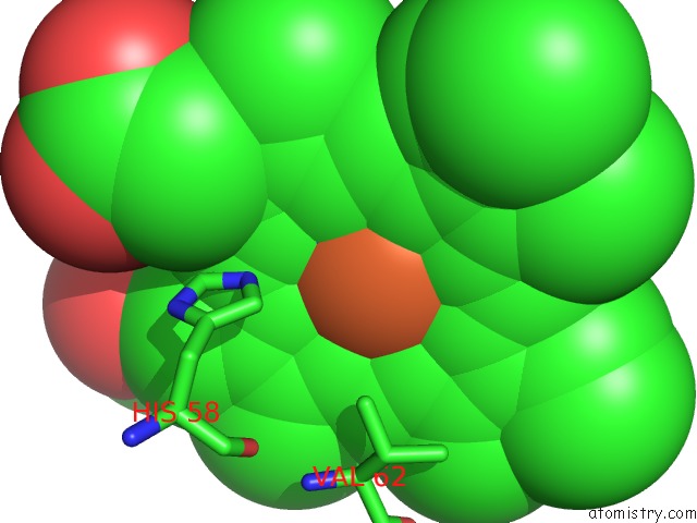
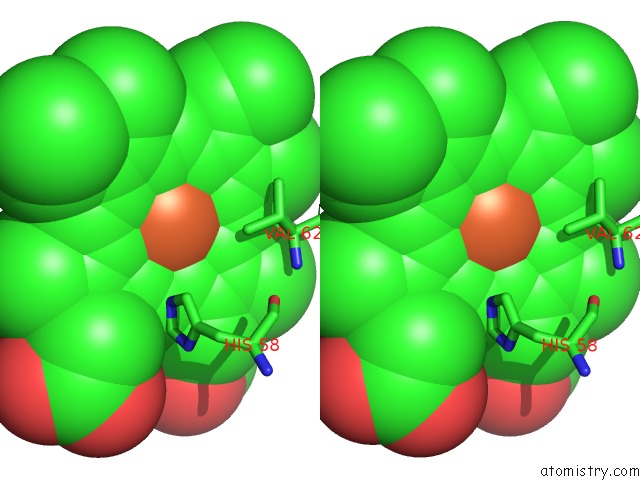
Iron binding site 1 out of 4 in 6kav
Go back to
Iron binding site 1 out
of 4 in the Carbonmonoxy Human Hemoglobin A in the R2 Quaternary Structure at 140 K: Light
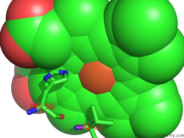
Mono view
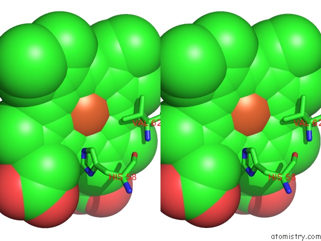
Stereo pair view
Mono view
Stereo pair view
A full contact list of Iron with other atoms in the Fe binding
site number 1 of Carbonmonoxy Human Hemoglobin A in the R2 Quaternary Structure at 140 K: Light within 5.0Å range:
|
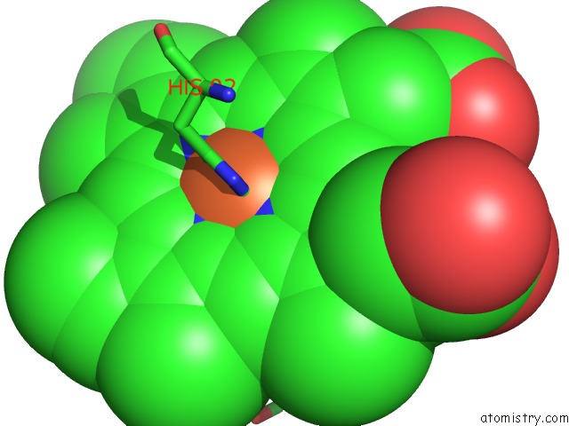
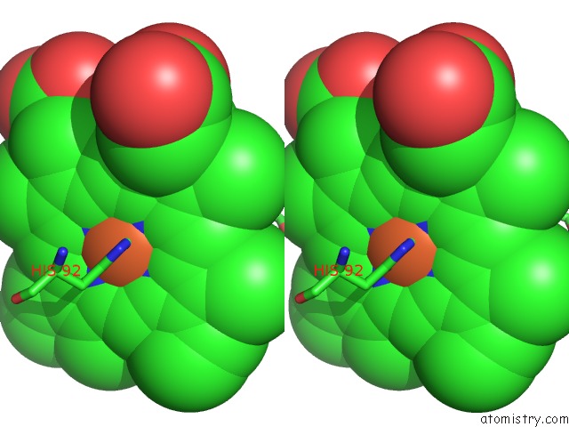
Iron binding site 2 out of 4 in 6kav
Go back to
Iron binding site 2 out
of 4 in the Carbonmonoxy Human Hemoglobin A in the R2 Quaternary Structure at 140 K: Light
Mono view
Stereo pair view
Mono view
Stereo pair view
A full contact list of Iron with other atoms in the Fe binding
site number 2 of Carbonmonoxy Human Hemoglobin A in the R2 Quaternary Structure at 140 K: Light within 5.0Å range:
|
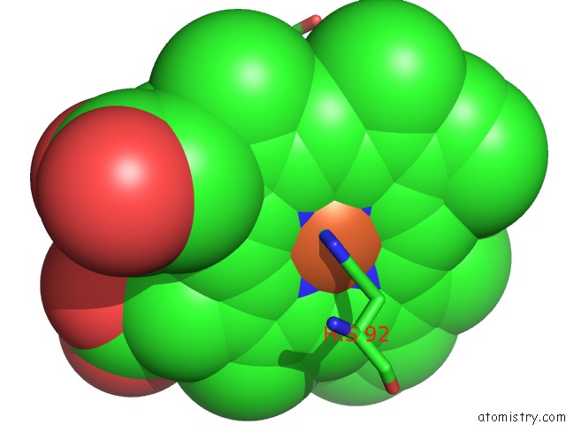
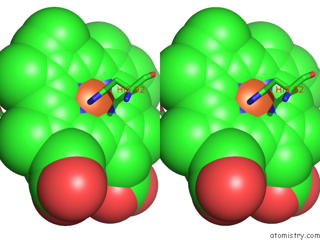
Iron binding site 3 out of 4 in 6kav
Go back to
Iron binding site 3 out
of 4 in the Carbonmonoxy Human Hemoglobin A in the R2 Quaternary Structure at 140 K: Light
Mono view
Stereo pair view
Mono view
Stereo pair view
A full contact list of Iron with other atoms in the Fe binding
site number 3 of Carbonmonoxy Human Hemoglobin A in the R2 Quaternary Structure at 140 K: Light within 5.0Å range:
|
Iron binding site 4 out of 4 in 6kav
Go back to
Iron binding site 4 out
of 4 in the Carbonmonoxy Human Hemoglobin A in the R2 Quaternary Structure at 140 K: Light
Mono view
Stereo pair view
Mono view
Stereo pair view
A full contact list of Iron with other atoms in the Fe binding
site number 4 of Carbonmonoxy Human Hemoglobin A in the R2 Quaternary Structure at 140 K: Light within 5.0Å range:
|
Reference:
N.Shibayama,
A.Sato-Tomita,
M.Ohki,
K.Ichiyanagi,
S.Y.Park.
Direct Observation of Ligand Migration Within Human Hemoglobin at Work Proc.Natl.Acad.Sci.Usa 2020.
ISSN: ESSN 1091-6490
Page generated: Tue Aug 6 23:57:20 2024
ISSN: ESSN 1091-6490
Last articles
Zn in 9J0NZn in 9J0O
Zn in 9J0P
Zn in 9FJX
Zn in 9EKB
Zn in 9C0F
Zn in 9CAH
Zn in 9CH0
Zn in 9CH3
Zn in 9CH1