Iron »
PDB 6l3a-6ll0 »
6l69 »
Iron in PDB 6l69: Crystal Structure of CYP154C2 From Streptomyces Avermitilis
Protein crystallography data
The structure of Crystal Structure of CYP154C2 From Streptomyces Avermitilis, PDB code: 6l69
was solved by
L.H.Xu,
S.Fushinobu,
with X-Ray Crystallography technique. A brief refinement statistics is given in the table below:
| Resolution Low / High (Å) | 33.89 / 1.50 |
| Space group | P 1 21 1 |
| Cell size a, b, c (Å), α, β, γ (°) | 67.780, 53.390, 98.581, 90.00, 90.04, 90.00 |
| R / Rfree (%) | 16.8 / 19.3 |
Iron Binding Sites:
The binding sites of Iron atom in the Crystal Structure of CYP154C2 From Streptomyces Avermitilis
(pdb code 6l69). This binding sites where shown within
5.0 Angstroms radius around Iron atom.
In total 2 binding sites of Iron where determined in the Crystal Structure of CYP154C2 From Streptomyces Avermitilis, PDB code: 6l69:
Jump to Iron binding site number: 1; 2;
In total 2 binding sites of Iron where determined in the Crystal Structure of CYP154C2 From Streptomyces Avermitilis, PDB code: 6l69:
Jump to Iron binding site number: 1; 2;
Iron binding site 1 out of 2 in 6l69
Go back to
Iron binding site 1 out
of 2 in the Crystal Structure of CYP154C2 From Streptomyces Avermitilis
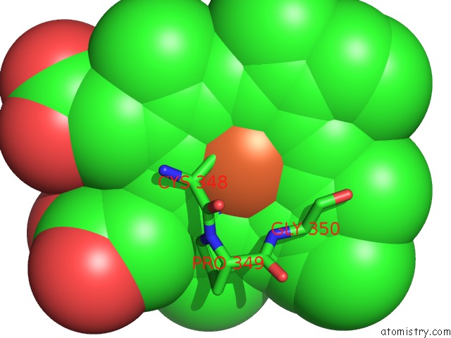
Mono view
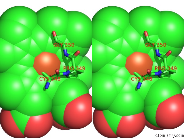
Stereo pair view
Mono view
Stereo pair view
A full contact list of Iron with other atoms in the Fe binding
site number 1 of Crystal Structure of CYP154C2 From Streptomyces Avermitilis within 5.0Å range:
|
Iron binding site 2 out of 2 in 6l69
Go back to
Iron binding site 2 out
of 2 in the Crystal Structure of CYP154C2 From Streptomyces Avermitilis
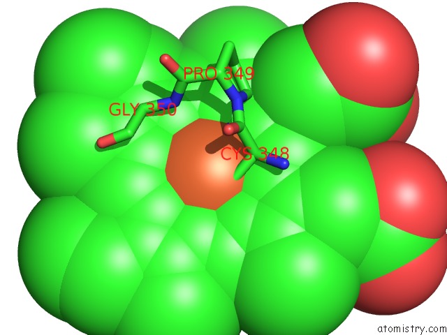
Mono view
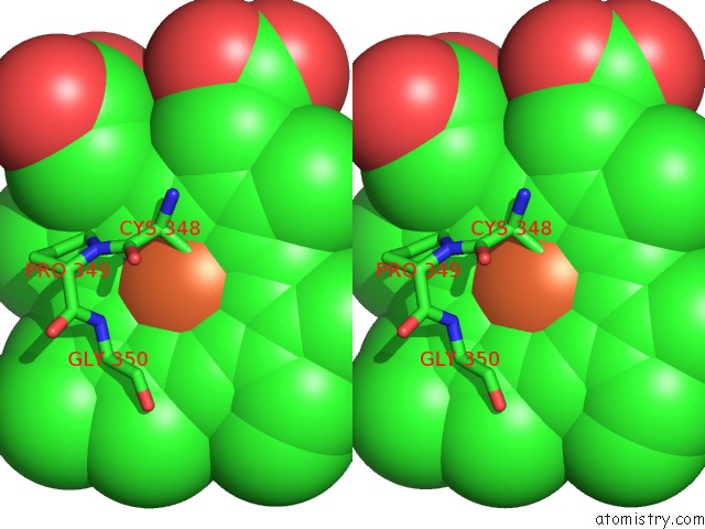
Stereo pair view
Mono view
Stereo pair view
A full contact list of Iron with other atoms in the Fe binding
site number 2 of Crystal Structure of CYP154C2 From Streptomyces Avermitilis within 5.0Å range:
|
Reference:
Q.Wang,
B.Ma,
S.Fushinobu,
C.Zhang,
L.H.Xu.
Regio- and Stereoselective Hydroxylation of Testosterone By A Novel Cytochrome P450 154C2 From Streptomyces Avermitilis. Biochem.Biophys.Res.Commun. V. 522 355 2020.
ISSN: ESSN 1090-2104
PubMed: 31767148
DOI: 10.1016/J.BBRC.2019.11.091
Page generated: Wed Aug 7 00:42:50 2024
ISSN: ESSN 1090-2104
PubMed: 31767148
DOI: 10.1016/J.BBRC.2019.11.091
Last articles
Zn in 9MJ5Zn in 9HNW
Zn in 9G0L
Zn in 9FNE
Zn in 9DZN
Zn in 9E0I
Zn in 9D32
Zn in 9DAK
Zn in 8ZXC
Zn in 8ZUF