Iron »
PDB 6ll4-6m7e »
6m1w »
Iron in PDB 6m1w: Structure of the 2-Aminoisobutyric Acid Monooxygenase Hydroxylase
Protein crystallography data
The structure of Structure of the 2-Aminoisobutyric Acid Monooxygenase Hydroxylase, PDB code: 6m1w
was solved by
M.Hibi,
B.Mikami,
J.Ogawa,
with X-Ray Crystallography technique. A brief refinement statistics is given in the table below:
| Resolution Low / High (Å) | 45.12 / 2.75 |
| Space group | C 2 2 21 |
| Cell size a, b, c (Å), α, β, γ (°) | 125.49, 208.05, 90.66, 90, 90, 90 |
| R / Rfree (%) | 19 / 25 |
Other elements in 6m1w:
The structure of Structure of the 2-Aminoisobutyric Acid Monooxygenase Hydroxylase also contains other interesting chemical elements:
| Chlorine | (Cl) | 1 atom |
| Zinc | (Zn) | 1 atom |
Iron Binding Sites:
The binding sites of Iron atom in the Structure of the 2-Aminoisobutyric Acid Monooxygenase Hydroxylase
(pdb code 6m1w). This binding sites where shown within
5.0 Angstroms radius around Iron atom.
In total 2 binding sites of Iron where determined in the Structure of the 2-Aminoisobutyric Acid Monooxygenase Hydroxylase, PDB code: 6m1w:
Jump to Iron binding site number: 1; 2;
In total 2 binding sites of Iron where determined in the Structure of the 2-Aminoisobutyric Acid Monooxygenase Hydroxylase, PDB code: 6m1w:
Jump to Iron binding site number: 1; 2;
Iron binding site 1 out of 2 in 6m1w
Go back to
Iron binding site 1 out
of 2 in the Structure of the 2-Aminoisobutyric Acid Monooxygenase Hydroxylase
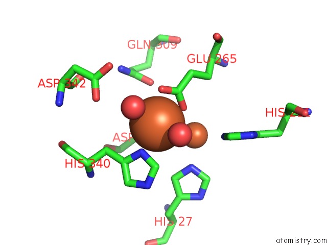
Mono view
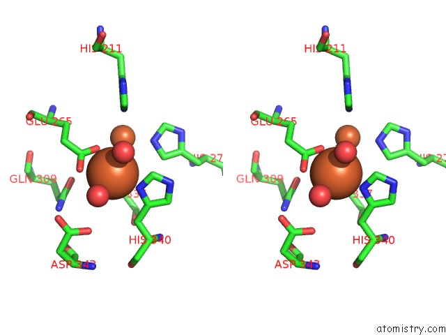
Stereo pair view
Mono view
Stereo pair view
A full contact list of Iron with other atoms in the Fe binding
site number 1 of Structure of the 2-Aminoisobutyric Acid Monooxygenase Hydroxylase within 5.0Å range:
|
Iron binding site 2 out of 2 in 6m1w
Go back to
Iron binding site 2 out
of 2 in the Structure of the 2-Aminoisobutyric Acid Monooxygenase Hydroxylase
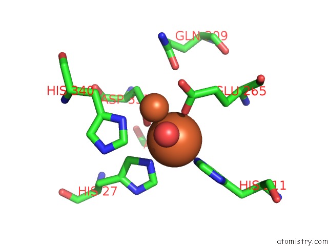
Mono view

Stereo pair view
Mono view
Stereo pair view
A full contact list of Iron with other atoms in the Fe binding
site number 2 of Structure of the 2-Aminoisobutyric Acid Monooxygenase Hydroxylase within 5.0Å range:
|
Reference:
M.Hibi,
B.Mikami,
J.Ogawa.
Catabolism of 2-Aminoisobutyric Acid Initiated By A Novel Four-Component Hydroxylase in Rhodococcus Wratislaviensis To Be Published.
Page generated: Wed Aug 6 09:44:05 2025
Last articles
Fe in 6Q6QFe in 6Q6P
Fe in 6Q3E
Fe in 6Q3D
Fe in 6Q5E
Fe in 6Q2Q
Fe in 6Q34
Fe in 6Q2P
Fe in 6Q31
Fe in 6Q2U