Iron »
PDB 6ll4-6m7e »
6m35 »
Iron in PDB 6m35: Crystal Structure of Sulfur Oxygenase Reductase From Sulfurisphaera Tokodaii
Enzymatic activity of Crystal Structure of Sulfur Oxygenase Reductase From Sulfurisphaera Tokodaii
All present enzymatic activity of Crystal Structure of Sulfur Oxygenase Reductase From Sulfurisphaera Tokodaii:
1.13.11.55;
1.13.11.55;
Protein crystallography data
The structure of Crystal Structure of Sulfur Oxygenase Reductase From Sulfurisphaera Tokodaii, PDB code: 6m35
was solved by
Y.Sato,
T.Yabuki,
T.Arakawa,
C.Yamada,
S.Fushinobu,
T.Wakagi,
with X-Ray Crystallography technique. A brief refinement statistics is given in the table below:
| Resolution Low / High (Å) | 48.68 / 1.73 |
| Space group | I 2 3 1 |
| Cell size a, b, c (Å), α, β, γ (°) | 299.806, 299.806, 299.806, 90.00, 90.00, 90.00 |
| R / Rfree (%) | 16.2 / 17.9 |
Iron Binding Sites:
The binding sites of Iron atom in the Crystal Structure of Sulfur Oxygenase Reductase From Sulfurisphaera Tokodaii
(pdb code 6m35). This binding sites where shown within
5.0 Angstroms radius around Iron atom.
In total 8 binding sites of Iron where determined in the Crystal Structure of Sulfur Oxygenase Reductase From Sulfurisphaera Tokodaii, PDB code: 6m35:
Jump to Iron binding site number: 1; 2; 3; 4; 5; 6; 7; 8;
In total 8 binding sites of Iron where determined in the Crystal Structure of Sulfur Oxygenase Reductase From Sulfurisphaera Tokodaii, PDB code: 6m35:
Jump to Iron binding site number: 1; 2; 3; 4; 5; 6; 7; 8;
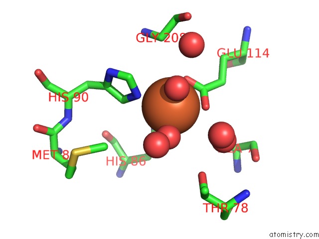
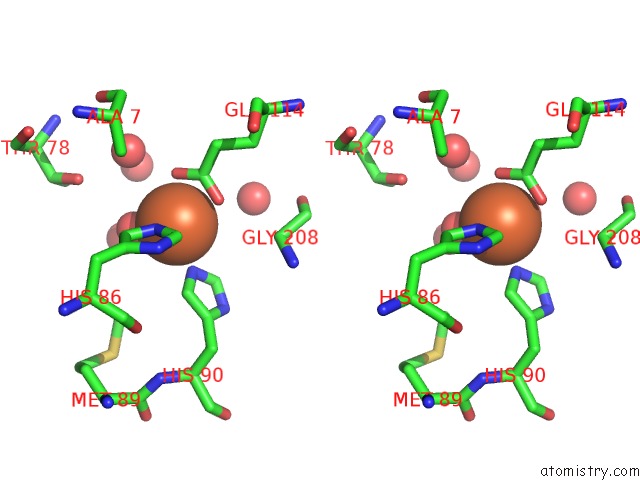
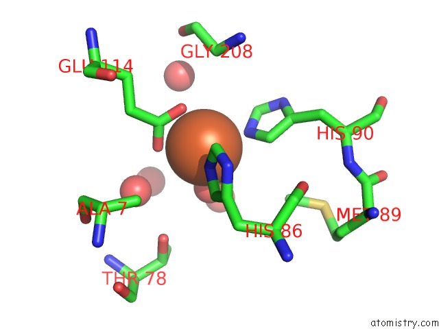
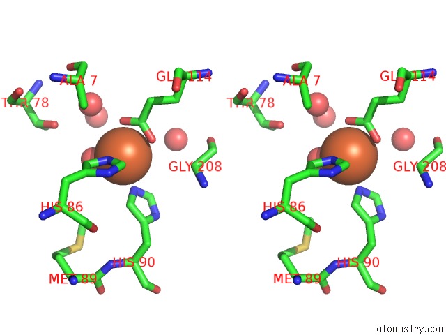
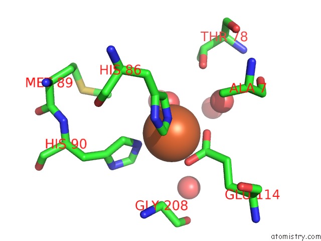
Iron binding site 1 out of 8 in 6m35
Go back to
Iron binding site 1 out
of 8 in the Crystal Structure of Sulfur Oxygenase Reductase From Sulfurisphaera Tokodaii
Mono view
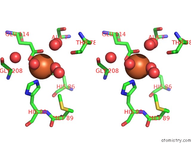
Stereo pair view
Mono view
Stereo pair view
A full contact list of Iron with other atoms in the Fe binding
site number 1 of Crystal Structure of Sulfur Oxygenase Reductase From Sulfurisphaera Tokodaii within 5.0Å range:
|
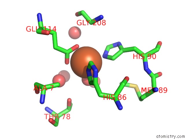
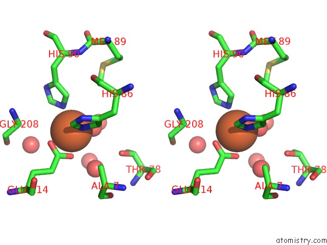
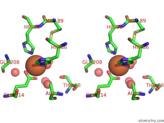
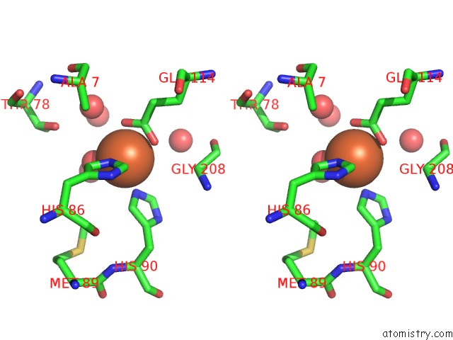
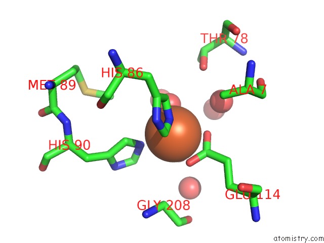
Iron binding site 2 out of 8 in 6m35
Go back to
Iron binding site 2 out
of 8 in the Crystal Structure of Sulfur Oxygenase Reductase From Sulfurisphaera Tokodaii
Mono view

Stereo pair view
Mono view
Stereo pair view
A full contact list of Iron with other atoms in the Fe binding
site number 2 of Crystal Structure of Sulfur Oxygenase Reductase From Sulfurisphaera Tokodaii within 5.0Å range:
|
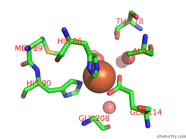
Iron binding site 3 out of 8 in 6m35
Go back to
Iron binding site 3 out
of 8 in the Crystal Structure of Sulfur Oxygenase Reductase From Sulfurisphaera Tokodaii
Mono view
Stereo pair view
Mono view
Stereo pair view
A full contact list of Iron with other atoms in the Fe binding
site number 3 of Crystal Structure of Sulfur Oxygenase Reductase From Sulfurisphaera Tokodaii within 5.0Å range:
|
Iron binding site 4 out of 8 in 6m35
Go back to
Iron binding site 4 out
of 8 in the Crystal Structure of Sulfur Oxygenase Reductase From Sulfurisphaera Tokodaii
Mono view
Stereo pair view
Mono view
Stereo pair view
A full contact list of Iron with other atoms in the Fe binding
site number 4 of Crystal Structure of Sulfur Oxygenase Reductase From Sulfurisphaera Tokodaii within 5.0Å range:
|
Iron binding site 5 out of 8 in 6m35
Go back to
Iron binding site 5 out
of 8 in the Crystal Structure of Sulfur Oxygenase Reductase From Sulfurisphaera Tokodaii

Mono view
Stereo pair view
Mono view
Stereo pair view
A full contact list of Iron with other atoms in the Fe binding
site number 5 of Crystal Structure of Sulfur Oxygenase Reductase From Sulfurisphaera Tokodaii within 5.0Å range:
|
Iron binding site 6 out of 8 in 6m35
Go back to
Iron binding site 6 out
of 8 in the Crystal Structure of Sulfur Oxygenase Reductase From Sulfurisphaera Tokodaii

Mono view
Stereo pair view
Mono view
Stereo pair view
A full contact list of Iron with other atoms in the Fe binding
site number 6 of Crystal Structure of Sulfur Oxygenase Reductase From Sulfurisphaera Tokodaii within 5.0Å range:
|
Iron binding site 7 out of 8 in 6m35
Go back to
Iron binding site 7 out
of 8 in the Crystal Structure of Sulfur Oxygenase Reductase From Sulfurisphaera Tokodaii
Mono view
Stereo pair view
Mono view
Stereo pair view
A full contact list of Iron with other atoms in the Fe binding
site number 7 of Crystal Structure of Sulfur Oxygenase Reductase From Sulfurisphaera Tokodaii within 5.0Å range:
|
Iron binding site 8 out of 8 in 6m35
Go back to
Iron binding site 8 out
of 8 in the Crystal Structure of Sulfur Oxygenase Reductase From Sulfurisphaera Tokodaii
Mono view

Stereo pair view
Mono view
Stereo pair view
A full contact list of Iron with other atoms in the Fe binding
site number 8 of Crystal Structure of Sulfur Oxygenase Reductase From Sulfurisphaera Tokodaii within 5.0Å range:
|
Reference:
Y.Sato,
T.Yabuki,
N.Adachi,
T.Moriya,
T.Arakawa,
M.Kawasaki,
C.Yamada,
T.Senda,
S.Fushinobu,
T.Wakagi.
Crystallographic and Cryogenic Electron Microscopic Structures and Enzymatic Characterization of Sulfur Oxygenase Reductase Fromsulfurisphaera Tokodaii. J Struct Biol X V. 4 00030 2020.
ISSN: ESSN 2590-1524
PubMed: 32775998
DOI: 10.1016/J.YJSBX.2020.100030
Page generated: Wed Aug 6 09:47:42 2025
ISSN: ESSN 2590-1524
PubMed: 32775998
DOI: 10.1016/J.YJSBX.2020.100030
Last articles
Fe in 6Q6QFe in 6Q6P
Fe in 6Q3E
Fe in 6Q3D
Fe in 6Q5E
Fe in 6Q2Q
Fe in 6Q34
Fe in 6Q2P
Fe in 6Q31
Fe in 6Q2U