Iron »
PDB 6m7e-6n1y »
6mi0 »
Iron in PDB 6mi0: Crystal Structure of the P450 Domain of the CYP51-Ferredoxin Fusion Protein From Methylococcus Capsulatus, Ligand-Free State
Enzymatic activity of Crystal Structure of the P450 Domain of the CYP51-Ferredoxin Fusion Protein From Methylococcus Capsulatus, Ligand-Free State
All present enzymatic activity of Crystal Structure of the P450 Domain of the CYP51-Ferredoxin Fusion Protein From Methylococcus Capsulatus, Ligand-Free State:
1.14.13.70;
1.14.13.70;
Protein crystallography data
The structure of Crystal Structure of the P450 Domain of the CYP51-Ferredoxin Fusion Protein From Methylococcus Capsulatus, Ligand-Free State, PDB code: 6mi0
was solved by
T.Hargrove,
Z.Wawrzak,
D.C.Lamb,
G.I.Lepesheva,
with X-Ray Crystallography technique. A brief refinement statistics is given in the table below:
| Resolution Low / High (Å) | 65.84 / 2.73 |
| Space group | P 31 2 1 |
| Cell size a, b, c (Å), α, β, γ (°) | 151.861, 151.861, 67.246, 90.00, 90.00, 120.00 |
| R / Rfree (%) | 21.4 / 23.4 |
Iron Binding Sites:
The binding sites of Iron atom in the Crystal Structure of the P450 Domain of the CYP51-Ferredoxin Fusion Protein From Methylococcus Capsulatus, Ligand-Free State
(pdb code 6mi0). This binding sites where shown within
5.0 Angstroms radius around Iron atom.
In total only one binding site of Iron was determined in the Crystal Structure of the P450 Domain of the CYP51-Ferredoxin Fusion Protein From Methylococcus Capsulatus, Ligand-Free State, PDB code: 6mi0:
In total only one binding site of Iron was determined in the Crystal Structure of the P450 Domain of the CYP51-Ferredoxin Fusion Protein From Methylococcus Capsulatus, Ligand-Free State, PDB code: 6mi0:
Iron binding site 1 out of 1 in 6mi0
Go back to
Iron binding site 1 out
of 1 in the Crystal Structure of the P450 Domain of the CYP51-Ferredoxin Fusion Protein From Methylococcus Capsulatus, Ligand-Free State
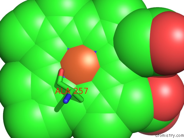
Mono view
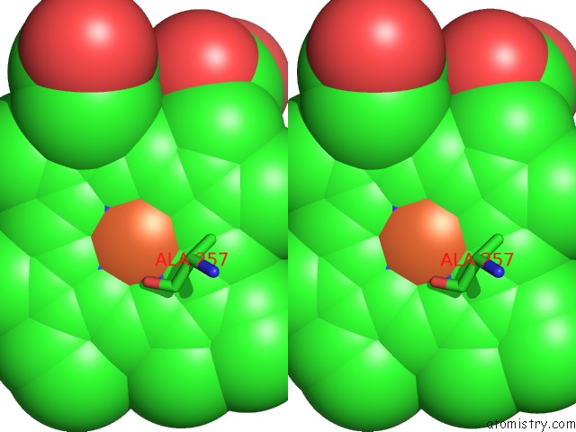
Stereo pair view
Mono view
Stereo pair view
A full contact list of Iron with other atoms in the Fe binding
site number 1 of Crystal Structure of the P450 Domain of the CYP51-Ferredoxin Fusion Protein From Methylococcus Capsulatus, Ligand-Free State within 5.0Å range:
|
Reference:
T.Hargrove,
Z.Wawrzak,
D.C.Lamb,
G.I.Lepesheva.
Crystal Structure of the P450 Domain of the CYP51-Ferredoxin Fusion Protein From Methylococcus Capsulatus To Be Published.
Page generated: Wed Aug 7 02:23:58 2024
Last articles
Zn in 9JYWZn in 9IR4
Zn in 9IR3
Zn in 9GMX
Zn in 9GMW
Zn in 9JEJ
Zn in 9ERF
Zn in 9ERE
Zn in 9EGV
Zn in 9EGW