Iron »
PDB 6m7e-6n1y »
6mp9 »
Iron in PDB 6mp9: X-Ray Crystal Structure of Vioc Bound to Fe(II), 2-Oxo-5- Guanidinopentanoic Acid, and Succinate
Enzymatic activity of X-Ray Crystal Structure of Vioc Bound to Fe(II), 2-Oxo-5- Guanidinopentanoic Acid, and Succinate
All present enzymatic activity of X-Ray Crystal Structure of Vioc Bound to Fe(II), 2-Oxo-5- Guanidinopentanoic Acid, and Succinate:
1.14.11.41;
1.14.11.41;
Protein crystallography data
The structure of X-Ray Crystal Structure of Vioc Bound to Fe(II), 2-Oxo-5- Guanidinopentanoic Acid, and Succinate, PDB code: 6mp9
was solved by
N.P.Dunham,
A.K.Boal,
with X-Ray Crystallography technique. A brief refinement statistics is given in the table below:
| Resolution Low / High (Å) | 59.39 / 1.89 |
| Space group | C 1 2 1 |
| Cell size a, b, c (Å), α, β, γ (°) | 80.676, 66.940, 62.856, 90.00, 109.12, 90.00 |
| R / Rfree (%) | 19.3 / 21.2 |
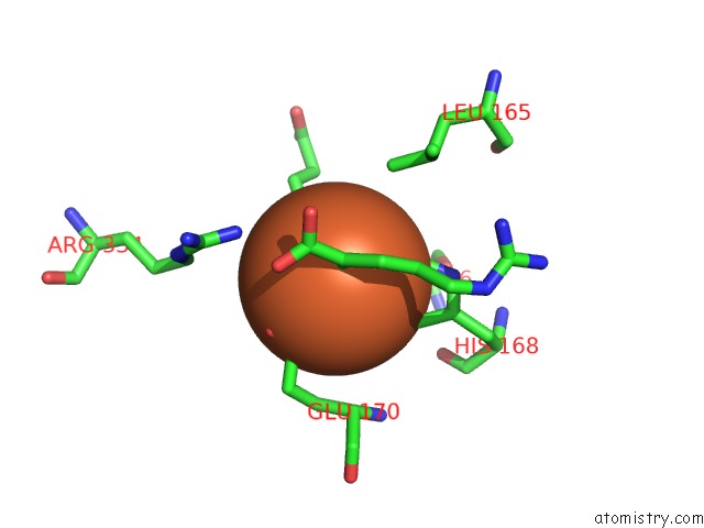
Iron Binding Sites:
The binding sites of Iron atom in the X-Ray Crystal Structure of Vioc Bound to Fe(II), 2-Oxo-5- Guanidinopentanoic Acid, and Succinate
(pdb code 6mp9). This binding sites where shown within
5.0 Angstroms radius around Iron atom.
In total only one binding site of Iron was determined in the X-Ray Crystal Structure of Vioc Bound to Fe(II), 2-Oxo-5- Guanidinopentanoic Acid, and Succinate, PDB code: 6mp9:
In total only one binding site of Iron was determined in the X-Ray Crystal Structure of Vioc Bound to Fe(II), 2-Oxo-5- Guanidinopentanoic Acid, and Succinate, PDB code: 6mp9:
Iron binding site 1 out of 1 in 6mp9
Go back to
Iron binding site 1 out
of 1 in the X-Ray Crystal Structure of Vioc Bound to Fe(II), 2-Oxo-5- Guanidinopentanoic Acid, and Succinate
Mono view
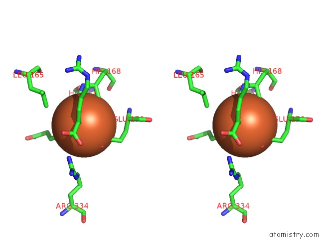
Stereo pair view
Mono view
Stereo pair view
A full contact list of Iron with other atoms in the Fe binding
site number 1 of X-Ray Crystal Structure of Vioc Bound to Fe(II), 2-Oxo-5- Guanidinopentanoic Acid, and Succinate within 5.0Å range:
|
Reference:
N.P.Dunham,
A.J.Mitchell,
J.M.Del Rio Pantoja,
C.Krebs,
J.M.Bollinger Jr.,
A.K.Boal.
Alpha-Amine Desaturation of D-Arginine By the Iron(II)- and 2-(Oxo)Glutarate-Dependent L-Arginine 3-Hydroxylase, Vioc. Biochemistry V. 57 6479 2018.
ISSN: ISSN 1520-4995
PubMed: 30403469
DOI: 10.1021/ACS.BIOCHEM.8B00901
Page generated: Wed Aug 7 02:25:20 2024
ISSN: ISSN 1520-4995
PubMed: 30403469
DOI: 10.1021/ACS.BIOCHEM.8B00901
Last articles
Zn in 9MJ5Zn in 9HNW
Zn in 9G0L
Zn in 9FNE
Zn in 9DZN
Zn in 9E0I
Zn in 9D32
Zn in 9DAK
Zn in 8ZXC
Zn in 8ZUF