Iron in PDB 6npa: X-Ray Crystal Structure of Tmpb, (R)-1-Hydroxy-2- Trimethylaminoethylphosphonate Oxygenase, with (R)-1-Hydroxy-2- Trimethylaminoethylphosphonate
Protein crystallography data
The structure of X-Ray Crystal Structure of Tmpb, (R)-1-Hydroxy-2- Trimethylaminoethylphosphonate Oxygenase, with (R)-1-Hydroxy-2- Trimethylaminoethylphosphonate, PDB code: 6npa
was solved by
L.J.Rajakovich,
A.J.Mitchell,
A.K.Boal,
with X-Ray Crystallography technique. A brief refinement statistics is given in the table below:
Resolution Low / High (Å)
|
50.00 /
1.73
|
Space group
|
C 2 2 21
|
Cell size a, b, c (Å), α, β, γ (°)
|
70.110,
151.322,
135.833,
90.00,
90.00,
90.00
|
R / Rfree (%)
|
22.2 /
25.1
|
Iron Binding Sites:
The binding sites of Iron atom in the X-Ray Crystal Structure of Tmpb, (R)-1-Hydroxy-2- Trimethylaminoethylphosphonate Oxygenase, with (R)-1-Hydroxy-2- Trimethylaminoethylphosphonate
(pdb code 6npa). This binding sites where shown within
5.0 Angstroms radius around Iron atom.
In total 10 binding sites of Iron where determined in the
X-Ray Crystal Structure of Tmpb, (R)-1-Hydroxy-2- Trimethylaminoethylphosphonate Oxygenase, with (R)-1-Hydroxy-2- Trimethylaminoethylphosphonate, PDB code: 6npa:
Jump to Iron binding site number:
1;
2;
3;
4;
5;
6;
7;
8;
9;
10;
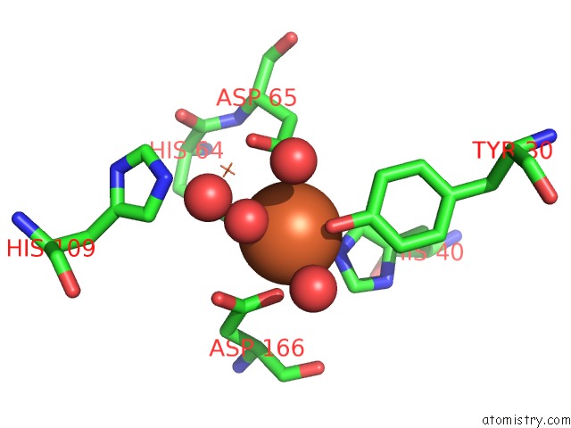
Iron binding site 1 out
of 10 in 6npa
Go back to
Iron Binding Sites List in 6npa
Iron binding site 1 out
of 10 in the X-Ray Crystal Structure of Tmpb, (R)-1-Hydroxy-2- Trimethylaminoethylphosphonate Oxygenase, with (R)-1-Hydroxy-2- Trimethylaminoethylphosphonate
Mono view
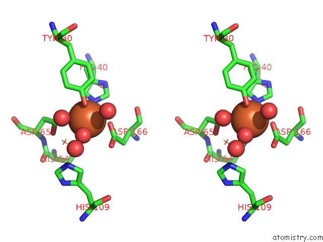
Stereo pair view
|
A full contact list of Iron with other atoms in the Fe binding
site number 1 of X-Ray Crystal Structure of Tmpb, (R)-1-Hydroxy-2- Trimethylaminoethylphosphonate Oxygenase, with (R)-1-Hydroxy-2- Trimethylaminoethylphosphonate within 5.0Å range:
probe
|
atom
|
residue
|
distance (Å)
|
B
|
Occ
|
A:Fe201
b:13.6
occ:1.00
|
O
|
A:HOH326
|
2.1
|
12.9
|
1.0
|
OD2
|
A:ASP65
|
2.1
|
12.6
|
1.0
|
OD1
|
A:ASP166
|
2.2
|
15.8
|
1.0
|
NE2
|
A:HIS40
|
2.2
|
13.7
|
1.0
|
OH
|
A:TYR30
|
2.2
|
17.1
|
1.0
|
NE2
|
A:HIS64
|
2.3
|
12.1
|
1.0
|
CG
|
A:ASP166
|
3.0
|
16.1
|
1.0
|
CG
|
A:ASP65
|
3.1
|
12.3
|
1.0
|
CD2
|
A:HIS40
|
3.1
|
13.5
|
1.0
|
CZ
|
A:TYR30
|
3.2
|
18.0
|
1.0
|
CD2
|
A:HIS64
|
3.2
|
12.2
|
1.0
|
CE1
|
A:HIS40
|
3.2
|
13.4
|
1.0
|
CE1
|
A:HIS64
|
3.2
|
11.9
|
1.0
|
OD1
|
A:ASP65
|
3.4
|
12.0
|
1.0
|
OD2
|
A:ASP166
|
3.4
|
16.7
|
1.0
|
FE
|
A:FE2202
|
3.6
|
15.3
|
1.0
|
O
|
A:HOH322
|
3.8
|
17.9
|
1.0
|
CE1
|
A:TYR30
|
3.9
|
18.3
|
1.0
|
O
|
A:HOH305
|
3.9
|
28.0
|
1.0
|
CE2
|
A:TYR30
|
3.9
|
18.1
|
1.0
|
CB
|
A:ASP166
|
4.2
|
16.1
|
1.0
|
CG
|
A:HIS40
|
4.3
|
13.3
|
1.0
|
ND1
|
A:HIS40
|
4.3
|
13.4
|
1.0
|
ND1
|
A:HIS64
|
4.3
|
12.1
|
1.0
|
CG
|
A:HIS64
|
4.3
|
11.9
|
1.0
|
CB
|
A:ASP65
|
4.3
|
12.6
|
1.0
|
CA
|
A:ASP166
|
4.6
|
16.0
|
1.0
|
NE2
|
A:HIS109
|
4.8
|
14.6
|
1.0
|
O
|
A:HOH360
|
4.9
|
16.3
|
1.0
|
O
|
A:ASP166
|
4.9
|
16.6
|
1.0
|
CD2
|
A:HIS109
|
4.9
|
14.4
|
1.0
|
|
Iron binding site 2 out
of 10 in 6npa
Go back to
Iron Binding Sites List in 6npa
Iron binding site 2 out
of 10 in the X-Ray Crystal Structure of Tmpb, (R)-1-Hydroxy-2- Trimethylaminoethylphosphonate Oxygenase, with (R)-1-Hydroxy-2- Trimethylaminoethylphosphonate
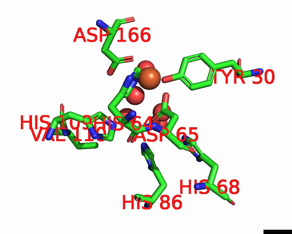
Mono view
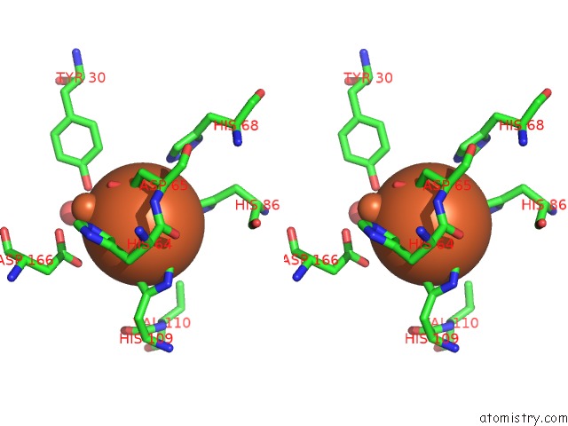
Stereo pair view
|
A full contact list of Iron with other atoms in the Fe binding
site number 2 of X-Ray Crystal Structure of Tmpb, (R)-1-Hydroxy-2- Trimethylaminoethylphosphonate Oxygenase, with (R)-1-Hydroxy-2- Trimethylaminoethylphosphonate within 5.0Å range:
probe
|
atom
|
residue
|
distance (Å)
|
B
|
Occ
|
A:Fe202
b:15.3
occ:1.00
|
O
|
A:HOH326
|
1.9
|
12.9
|
1.0
|
O
|
A:HOH360
|
2.2
|
16.3
|
1.0
|
NE2
|
A:HIS109
|
2.2
|
14.6
|
1.0
|
OD1
|
A:ASP65
|
2.2
|
12.0
|
1.0
|
O
|
A:HOH322
|
2.3
|
17.9
|
1.0
|
NE2
|
A:HIS86
|
2.3
|
18.4
|
1.0
|
CE1
|
A:HIS109
|
3.2
|
14.2
|
1.0
|
CD2
|
A:HIS109
|
3.2
|
14.4
|
1.0
|
CD2
|
A:HIS86
|
3.2
|
18.3
|
1.0
|
CG
|
A:ASP65
|
3.3
|
12.3
|
1.0
|
CE1
|
A:HIS86
|
3.4
|
18.6
|
1.0
|
OD2
|
A:ASP65
|
3.5
|
12.6
|
1.0
|
FE
|
A:FE201
|
3.6
|
13.6
|
1.0
|
OH
|
A:TYR30
|
3.9
|
17.1
|
1.0
|
CD2
|
A:HIS64
|
4.1
|
12.2
|
1.0
|
NE2
|
A:HIS68
|
4.2
|
20.5
|
1.0
|
ND1
|
A:HIS109
|
4.3
|
14.2
|
1.0
|
CG
|
A:HIS109
|
4.3
|
14.3
|
1.0
|
O
|
A:HOH371
|
4.4
|
29.4
|
1.0
|
OD2
|
A:ASP166
|
4.4
|
16.7
|
1.0
|
CG
|
A:HIS86
|
4.4
|
18.8
|
1.0
|
NE2
|
A:HIS64
|
4.4
|
12.1
|
1.0
|
ND1
|
A:HIS86
|
4.5
|
18.8
|
1.0
|
CB
|
A:ASP65
|
4.6
|
12.6
|
1.0
|
CG2
|
A:VAL110
|
4.9
|
16.4
|
1.0
|
O
|
A:HIS64
|
4.9
|
11.7
|
1.0
|
O
|
A:HOH305
|
4.9
|
28.0
|
1.0
|
OD1
|
A:ASP166
|
4.9
|
15.8
|
1.0
|
CD2
|
A:HIS68
|
4.9
|
20.7
|
1.0
|
CA
|
A:ASP65
|
5.0
|
12.9
|
1.0
|
|
Iron binding site 3 out
of 10 in 6npa
Go back to
Iron Binding Sites List in 6npa
Iron binding site 3 out
of 10 in the X-Ray Crystal Structure of Tmpb, (R)-1-Hydroxy-2- Trimethylaminoethylphosphonate Oxygenase, with (R)-1-Hydroxy-2- Trimethylaminoethylphosphonate
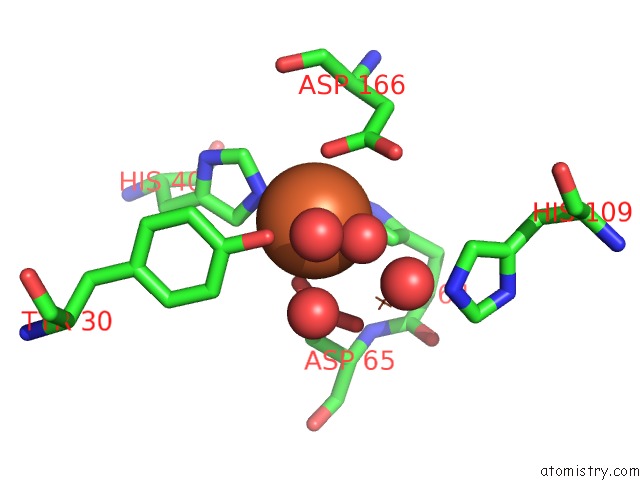
Mono view
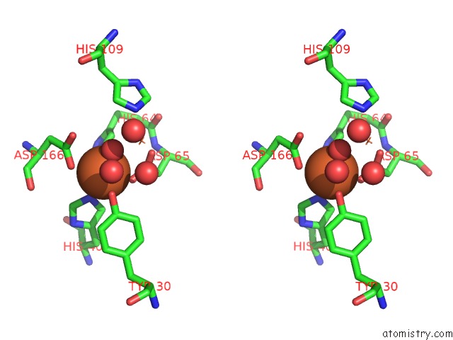
Stereo pair view
|
A full contact list of Iron with other atoms in the Fe binding
site number 3 of X-Ray Crystal Structure of Tmpb, (R)-1-Hydroxy-2- Trimethylaminoethylphosphonate Oxygenase, with (R)-1-Hydroxy-2- Trimethylaminoethylphosphonate within 5.0Å range:
probe
|
atom
|
residue
|
distance (Å)
|
B
|
Occ
|
B:Fe201
b:14.2
occ:1.00
|
O
|
B:HOH323
|
2.0
|
14.2
|
1.0
|
OD2
|
B:ASP65
|
2.1
|
12.9
|
1.0
|
NE2
|
B:HIS40
|
2.2
|
13.2
|
1.0
|
OD1
|
B:ASP166
|
2.2
|
16.5
|
1.0
|
OH
|
B:TYR30
|
2.3
|
16.3
|
1.0
|
NE2
|
B:HIS64
|
2.3
|
11.4
|
1.0
|
CG
|
B:ASP166
|
3.0
|
16.6
|
1.0
|
CG
|
B:ASP65
|
3.1
|
12.6
|
1.0
|
CD2
|
B:HIS40
|
3.2
|
13.1
|
1.0
|
CZ
|
B:TYR30
|
3.2
|
17.0
|
1.0
|
CD2
|
B:HIS64
|
3.2
|
11.5
|
1.0
|
CE1
|
B:HIS40
|
3.2
|
13.2
|
1.0
|
CE1
|
B:HIS64
|
3.3
|
11.4
|
1.0
|
OD2
|
B:ASP166
|
3.5
|
16.9
|
1.0
|
OD1
|
B:ASP65
|
3.5
|
11.7
|
1.0
|
FE
|
B:FE2204
|
3.6
|
14.5
|
1.0
|
O
|
B:HOH320
|
3.7
|
17.0
|
1.0
|
CE1
|
B:TYR30
|
3.9
|
17.5
|
1.0
|
CE2
|
B:TYR30
|
4.0
|
16.8
|
1.0
|
O
|
B:HOH339
|
4.1
|
23.0
|
1.0
|
CB
|
B:ASP166
|
4.2
|
16.7
|
1.0
|
ND1
|
B:HIS40
|
4.3
|
13.2
|
1.0
|
CG
|
B:HIS40
|
4.3
|
13.5
|
1.0
|
CB
|
B:ASP65
|
4.3
|
12.2
|
1.0
|
CG
|
B:HIS64
|
4.4
|
11.3
|
1.0
|
ND1
|
B:HIS64
|
4.4
|
11.5
|
1.0
|
CA
|
B:ASP166
|
4.6
|
16.6
|
1.0
|
NE2
|
B:HIS109
|
4.8
|
15.2
|
1.0
|
O
|
B:ASP166
|
4.9
|
17.8
|
1.0
|
CD2
|
B:HIS109
|
4.9
|
15.2
|
1.0
|
O
|
B:HOH362
|
4.9
|
14.0
|
1.0
|
|
Iron binding site 4 out
of 10 in 6npa
Go back to
Iron Binding Sites List in 6npa
Iron binding site 4 out
of 10 in the X-Ray Crystal Structure of Tmpb, (R)-1-Hydroxy-2- Trimethylaminoethylphosphonate Oxygenase, with (R)-1-Hydroxy-2- Trimethylaminoethylphosphonate
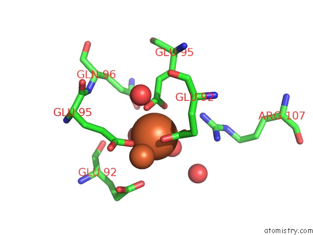
Mono view
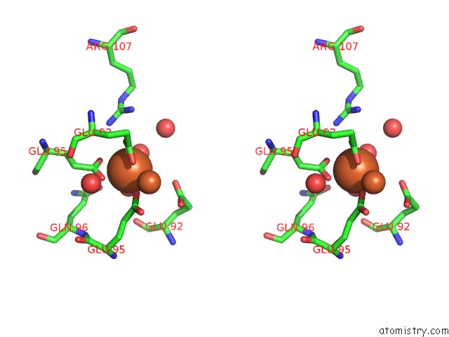
Stereo pair view
|
A full contact list of Iron with other atoms in the Fe binding
site number 4 of X-Ray Crystal Structure of Tmpb, (R)-1-Hydroxy-2- Trimethylaminoethylphosphonate Oxygenase, with (R)-1-Hydroxy-2- Trimethylaminoethylphosphonate within 5.0Å range:
probe
|
atom
|
residue
|
distance (Å)
|
B
|
Occ
|
B:Fe202
b:14.2
occ:0.50
|
OE2
|
A:GLU95
|
2.1
|
19.4
|
1.0
|
O
|
A:HOH344
|
2.2
|
21.8
|
1.0
|
O
|
B:HOH359
|
2.3
|
29.5
|
1.0
|
OE2
|
B:GLU95
|
2.3
|
20.3
|
1.0
|
OE2
|
B:GLU92
|
2.5
|
16.0
|
1.0
|
OE1
|
B:GLU92
|
2.6
|
17.2
|
1.0
|
OE1
|
B:GLU95
|
2.7
|
18.6
|
1.0
|
CD
|
B:GLU95
|
2.8
|
18.9
|
1.0
|
CD
|
B:GLU92
|
2.9
|
16.2
|
1.0
|
CD
|
A:GLU95
|
3.3
|
18.6
|
1.0
|
OE1
|
A:GLU92
|
3.8
|
24.5
|
1.0
|
FE
|
B:FE203
|
3.9
|
17.6
|
0.5
|
CG
|
A:GLU95
|
4.0
|
18.3
|
1.0
|
OE1
|
A:GLU95
|
4.2
|
19.3
|
1.0
|
NH2
|
B:ARG107
|
4.3
|
18.5
|
1.0
|
CG
|
B:GLU95
|
4.3
|
18.4
|
1.0
|
CG
|
B:GLU92
|
4.4
|
15.7
|
1.0
|
O
|
B:HOH307
|
4.5
|
17.7
|
1.0
|
CD
|
A:GLU92
|
4.6
|
22.7
|
1.0
|
O
|
B:HOH349
|
4.6
|
27.6
|
1.0
|
OE1
|
A:GLN96
|
4.7
|
29.9
|
1.0
|
NH1
|
B:ARG107
|
4.8
|
19.0
|
1.0
|
OE2
|
A:GLU92
|
4.8
|
23.8
|
1.0
|
O
|
A:GLU92
|
4.8
|
17.8
|
1.0
|
CZ
|
B:ARG107
|
5.0
|
17.9
|
1.0
|
|
Iron binding site 5 out
of 10 in 6npa
Go back to
Iron Binding Sites List in 6npa
Iron binding site 5 out
of 10 in the X-Ray Crystal Structure of Tmpb, (R)-1-Hydroxy-2- Trimethylaminoethylphosphonate Oxygenase, with (R)-1-Hydroxy-2- Trimethylaminoethylphosphonate
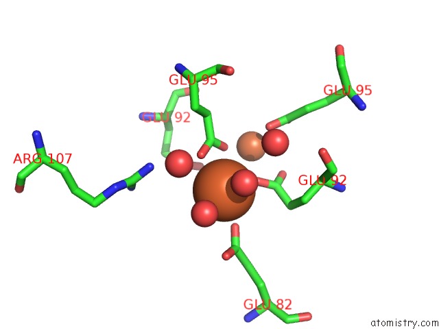
Mono view
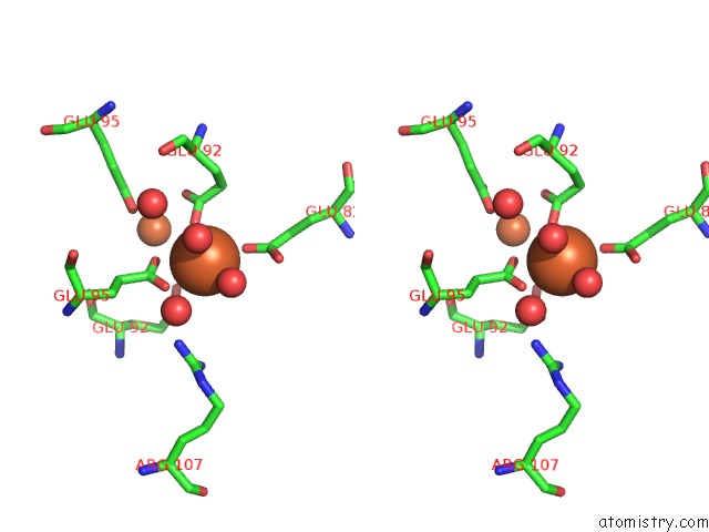
Stereo pair view
|
A full contact list of Iron with other atoms in the Fe binding
site number 5 of X-Ray Crystal Structure of Tmpb, (R)-1-Hydroxy-2- Trimethylaminoethylphosphonate Oxygenase, with (R)-1-Hydroxy-2- Trimethylaminoethylphosphonate within 5.0Å range:
probe
|
atom
|
residue
|
distance (Å)
|
B
|
Occ
|
B:Fe203
b:17.6
occ:0.50
|
OE1
|
B:GLU92
|
2.2
|
17.2
|
1.0
|
O
|
B:HOH333
|
2.3
|
40.1
|
1.0
|
OE2
|
B:GLU82
|
2.3
|
30.4
|
1.0
|
OE1
|
A:GLU95
|
2.5
|
19.3
|
1.0
|
O
|
B:HOH369
|
2.6
|
27.4
|
1.0
|
OE2
|
A:GLU95
|
2.7
|
19.4
|
1.0
|
CD
|
A:GLU95
|
3.0
|
18.6
|
1.0
|
CD
|
B:GLU92
|
3.3
|
16.2
|
1.0
|
CD
|
B:GLU82
|
3.4
|
32.9
|
1.0
|
OE1
|
B:GLU82
|
3.8
|
32.3
|
1.0
|
FE
|
B:FE202
|
3.9
|
14.2
|
0.5
|
OE2
|
A:GLU92
|
4.0
|
23.8
|
1.0
|
CG
|
B:GLU92
|
4.0
|
15.7
|
1.0
|
OE2
|
B:GLU92
|
4.3
|
16.0
|
1.0
|
O
|
A:HOH341
|
4.3
|
28.9
|
1.0
|
O
|
B:HOH307
|
4.3
|
17.7
|
1.0
|
NH2
|
A:ARG107
|
4.4
|
24.1
|
1.0
|
CG
|
A:GLU95
|
4.5
|
18.3
|
1.0
|
OE2
|
B:GLU95
|
4.5
|
20.3
|
1.0
|
OE1
|
A:GLU92
|
4.6
|
24.5
|
1.0
|
CB
|
B:GLU92
|
4.6
|
15.3
|
1.0
|
CG
|
B:GLU82
|
4.7
|
34.0
|
1.0
|
CD
|
A:GLU92
|
4.7
|
22.7
|
1.0
|
|
Iron binding site 6 out
of 10 in 6npa
Go back to
Iron Binding Sites List in 6npa
Iron binding site 6 out
of 10 in the X-Ray Crystal Structure of Tmpb, (R)-1-Hydroxy-2- Trimethylaminoethylphosphonate Oxygenase, with (R)-1-Hydroxy-2- Trimethylaminoethylphosphonate
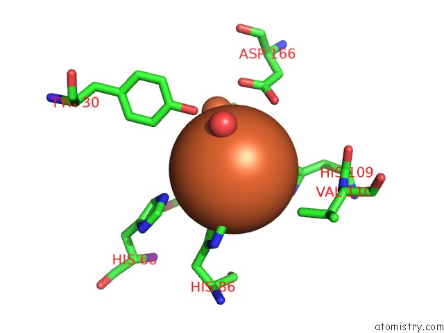
Mono view
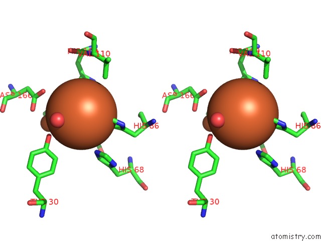
Stereo pair view
|
A full contact list of Iron with other atoms in the Fe binding
site number 6 of X-Ray Crystal Structure of Tmpb, (R)-1-Hydroxy-2- Trimethylaminoethylphosphonate Oxygenase, with (R)-1-Hydroxy-2- Trimethylaminoethylphosphonate within 5.0Å range:
probe
|
atom
|
residue
|
distance (Å)
|
B
|
Occ
|
B:Fe204
b:14.5
occ:1.00
|
O
|
B:HOH323
|
2.0
|
14.2
|
1.0
|
OD1
|
B:ASP65
|
2.2
|
11.7
|
1.0
|
O
|
B:HOH362
|
2.2
|
14.0
|
1.0
|
NE2
|
B:HIS109
|
2.3
|
15.2
|
1.0
|
O
|
B:HOH320
|
2.3
|
17.0
|
1.0
|
NE2
|
B:HIS86
|
2.3
|
18.1
|
1.0
|
CG
|
B:ASP65
|
3.2
|
12.6
|
1.0
|
CD2
|
B:HIS109
|
3.2
|
15.2
|
1.0
|
CE1
|
B:HIS109
|
3.3
|
15.3
|
1.0
|
CD2
|
B:HIS86
|
3.3
|
18.0
|
1.0
|
CE1
|
B:HIS86
|
3.3
|
18.2
|
1.0
|
OD2
|
B:ASP65
|
3.5
|
12.9
|
1.0
|
FE
|
B:FE201
|
3.6
|
14.2
|
1.0
|
O
|
B:HOH339
|
3.7
|
23.0
|
1.0
|
OH
|
B:TYR30
|
4.0
|
16.3
|
1.0
|
CD2
|
B:HIS64
|
4.1
|
11.5
|
1.0
|
NE2
|
B:HIS68
|
4.1
|
19.8
|
1.0
|
ND1
|
B:HIS109
|
4.4
|
15.2
|
1.0
|
CG
|
B:HIS109
|
4.4
|
15.1
|
1.0
|
ND1
|
B:HIS86
|
4.4
|
18.4
|
1.0
|
CG
|
B:HIS86
|
4.4
|
18.2
|
1.0
|
OD2
|
B:ASP166
|
4.5
|
16.9
|
1.0
|
NE2
|
B:HIS64
|
4.5
|
11.4
|
1.0
|
CB
|
B:ASP65
|
4.6
|
12.2
|
1.0
|
CG2
|
B:VAL110
|
4.8
|
15.7
|
1.0
|
O
|
B:HIS64
|
4.8
|
12.2
|
1.0
|
CE1
|
B:HIS68
|
4.9
|
20.2
|
1.0
|
CD2
|
B:HIS68
|
5.0
|
20.1
|
1.0
|
CA
|
B:ASP65
|
5.0
|
12.5
|
1.0
|
OD1
|
B:ASP166
|
5.0
|
16.5
|
1.0
|
|
Iron binding site 7 out
of 10 in 6npa
Go back to
Iron Binding Sites List in 6npa
Iron binding site 7 out
of 10 in the X-Ray Crystal Structure of Tmpb, (R)-1-Hydroxy-2- Trimethylaminoethylphosphonate Oxygenase, with (R)-1-Hydroxy-2- Trimethylaminoethylphosphonate
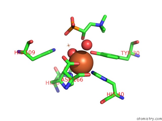
Mono view
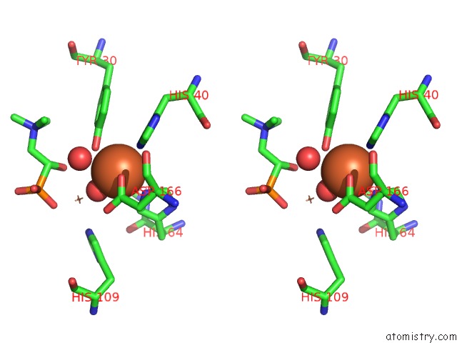
Stereo pair view
|
A full contact list of Iron with other atoms in the Fe binding
site number 7 of X-Ray Crystal Structure of Tmpb, (R)-1-Hydroxy-2- Trimethylaminoethylphosphonate Oxygenase, with (R)-1-Hydroxy-2- Trimethylaminoethylphosphonate within 5.0Å range:
probe
|
atom
|
residue
|
distance (Å)
|
B
|
Occ
|
C:Fe201
b:15.3
occ:1.00
|
O
|
C:HOH317
|
1.9
|
14.0
|
1.0
|
OD2
|
C:ASP65
|
2.1
|
15.5
|
1.0
|
OH
|
C:TYR30
|
2.2
|
22.2
|
1.0
|
NE2
|
C:HIS40
|
2.2
|
17.0
|
1.0
|
OD1
|
C:ASP166
|
2.3
|
16.8
|
1.0
|
NE2
|
C:HIS64
|
2.3
|
14.9
|
1.0
|
CG
|
C:ASP166
|
3.0
|
16.8
|
1.0
|
CG
|
C:ASP65
|
3.1
|
14.8
|
1.0
|
CD2
|
C:HIS64
|
3.2
|
14.9
|
1.0
|
CZ
|
C:TYR30
|
3.2
|
22.9
|
1.0
|
CD2
|
C:HIS40
|
3.2
|
16.9
|
1.0
|
CE1
|
C:HIS40
|
3.2
|
17.1
|
1.0
|
OD2
|
C:ASP166
|
3.3
|
17.4
|
1.0
|
CE1
|
C:HIS64
|
3.4
|
14.9
|
1.0
|
OD1
|
C:ASP65
|
3.5
|
15.4
|
1.0
|
O
|
C:HOH306
|
3.6
|
22.7
|
1.0
|
FE
|
C:FE2202
|
3.6
|
15.9
|
1.0
|
CE1
|
C:TYR30
|
3.9
|
23.1
|
1.0
|
O1
|
C:KVP203
|
3.9
|
24.4
|
1.0
|
CE2
|
C:TYR30
|
4.0
|
23.2
|
1.0
|
CB
|
C:ASP166
|
4.2
|
16.7
|
1.0
|
CB
|
C:ASP65
|
4.3
|
14.2
|
1.0
|
ND1
|
C:HIS40
|
4.3
|
17.6
|
1.0
|
CG
|
C:HIS40
|
4.3
|
17.3
|
1.0
|
CG
|
C:HIS64
|
4.3
|
14.4
|
1.0
|
ND1
|
C:HIS64
|
4.4
|
14.7
|
1.0
|
C1
|
C:KVP203
|
4.6
|
24.9
|
1.0
|
CA
|
C:ASP166
|
4.7
|
16.7
|
1.0
|
O11
|
C:KVP203
|
4.8
|
23.7
|
1.0
|
NE2
|
C:HIS109
|
4.8
|
16.6
|
1.0
|
O
|
C:ASP166
|
5.0
|
17.9
|
1.0
|
CD2
|
C:HIS109
|
5.0
|
16.1
|
1.0
|
|
Iron binding site 8 out
of 10 in 6npa
Go back to
Iron Binding Sites List in 6npa
Iron binding site 8 out
of 10 in the X-Ray Crystal Structure of Tmpb, (R)-1-Hydroxy-2- Trimethylaminoethylphosphonate Oxygenase, with (R)-1-Hydroxy-2- Trimethylaminoethylphosphonate
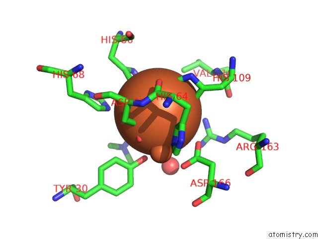
Mono view
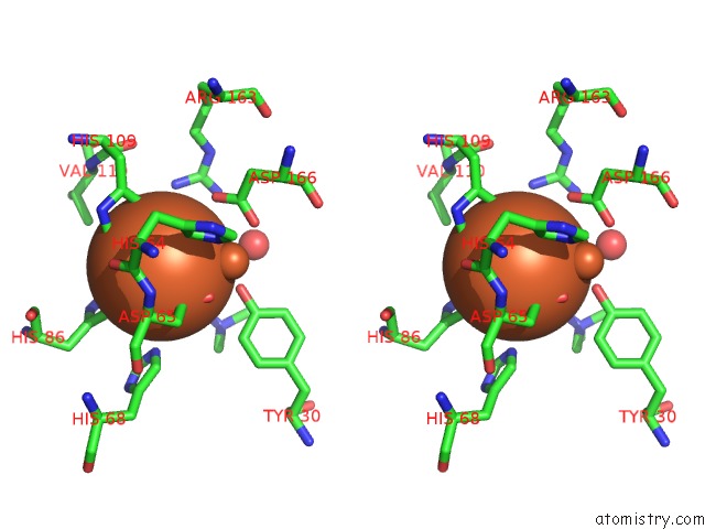
Stereo pair view
|
A full contact list of Iron with other atoms in the Fe binding
site number 8 of X-Ray Crystal Structure of Tmpb, (R)-1-Hydroxy-2- Trimethylaminoethylphosphonate Oxygenase, with (R)-1-Hydroxy-2- Trimethylaminoethylphosphonate within 5.0Å range:
probe
|
atom
|
residue
|
distance (Å)
|
B
|
Occ
|
C:Fe202
b:15.9
occ:1.00
|
O
|
C:HOH317
|
2.0
|
14.0
|
1.0
|
O11
|
C:KVP203
|
2.1
|
23.7
|
1.0
|
OD1
|
C:ASP65
|
2.2
|
15.4
|
1.0
|
NE2
|
C:HIS109
|
2.3
|
16.6
|
1.0
|
NE2
|
C:HIS86
|
2.3
|
19.3
|
1.0
|
O1
|
C:KVP203
|
2.4
|
24.4
|
1.0
|
CG
|
C:ASP65
|
3.2
|
14.8
|
1.0
|
CE1
|
C:HIS86
|
3.2
|
20.4
|
1.0
|
CD2
|
C:HIS109
|
3.2
|
16.1
|
1.0
|
P4
|
C:KVP203
|
3.2
|
24.3
|
1.0
|
CE1
|
C:HIS109
|
3.3
|
16.0
|
1.0
|
C1
|
C:KVP203
|
3.3
|
24.9
|
1.0
|
CD2
|
C:HIS86
|
3.3
|
19.5
|
1.0
|
OD2
|
C:ASP65
|
3.5
|
15.5
|
1.0
|
FE
|
C:FE201
|
3.6
|
15.3
|
1.0
|
OH
|
C:TYR30
|
4.0
|
22.2
|
1.0
|
O9
|
C:KVP203
|
4.1
|
24.4
|
1.0
|
NE2
|
C:HIS68
|
4.2
|
23.2
|
1.0
|
CD2
|
C:HIS64
|
4.2
|
14.9
|
1.0
|
OD2
|
C:ASP166
|
4.3
|
17.4
|
1.0
|
ND1
|
C:HIS86
|
4.4
|
20.2
|
1.0
|
O10
|
C:KVP203
|
4.4
|
25.3
|
1.0
|
ND1
|
C:HIS109
|
4.4
|
16.3
|
1.0
|
CG
|
C:HIS109
|
4.4
|
16.2
|
1.0
|
CG
|
C:HIS86
|
4.5
|
20.1
|
1.0
|
NH1
|
C:ARG163
|
4.5
|
15.8
|
1.0
|
CB
|
C:ASP65
|
4.5
|
14.2
|
1.0
|
NE2
|
C:HIS64
|
4.6
|
14.9
|
1.0
|
C2
|
C:KVP203
|
4.6
|
25.5
|
1.0
|
CD2
|
C:HIS68
|
4.9
|
22.4
|
1.0
|
CG2
|
C:VAL110
|
4.9
|
18.6
|
1.0
|
O
|
C:HOH306
|
4.9
|
22.7
|
1.0
|
O
|
C:HIS64
|
4.9
|
14.2
|
1.0
|
CA
|
C:ASP65
|
4.9
|
14.4
|
1.0
|
OD1
|
C:ASP166
|
5.0
|
16.8
|
1.0
|
|
Iron binding site 9 out
of 10 in 6npa
Go back to
Iron Binding Sites List in 6npa
Iron binding site 9 out
of 10 in the X-Ray Crystal Structure of Tmpb, (R)-1-Hydroxy-2- Trimethylaminoethylphosphonate Oxygenase, with (R)-1-Hydroxy-2- Trimethylaminoethylphosphonate
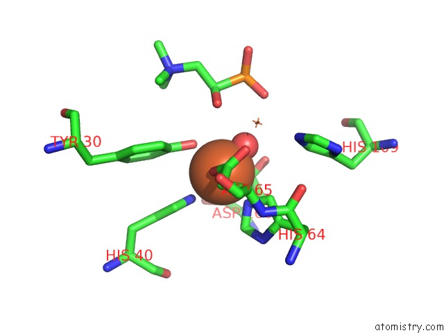
Mono view
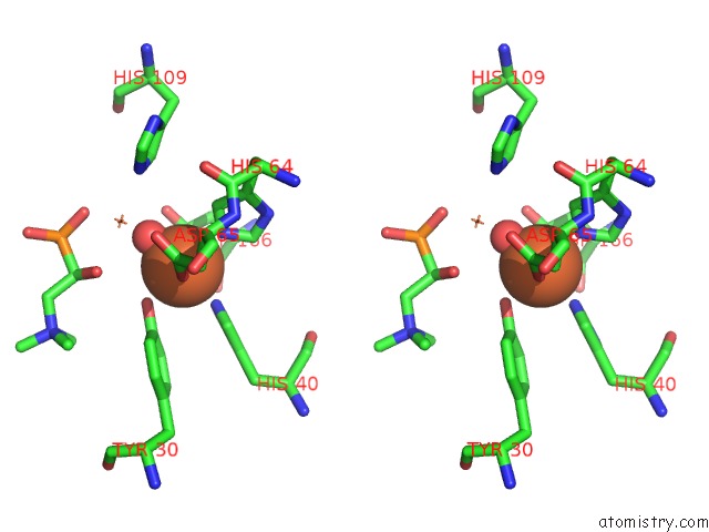
Stereo pair view
|
A full contact list of Iron with other atoms in the Fe binding
site number 9 of X-Ray Crystal Structure of Tmpb, (R)-1-Hydroxy-2- Trimethylaminoethylphosphonate Oxygenase, with (R)-1-Hydroxy-2- Trimethylaminoethylphosphonate within 5.0Å range:
probe
|
atom
|
residue
|
distance (Å)
|
B
|
Occ
|
D:Fe201
b:15.1
occ:1.00
|
O
|
D:HOH313
|
1.9
|
14.6
|
1.0
|
OD2
|
D:ASP65
|
2.0
|
14.6
|
1.0
|
OH
|
D:TYR30
|
2.2
|
22.2
|
1.0
|
NE2
|
D:HIS64
|
2.2
|
13.0
|
1.0
|
NE2
|
D:HIS40
|
2.3
|
16.5
|
1.0
|
OD1
|
D:ASP166
|
2.3
|
16.2
|
1.0
|
CG
|
D:ASP166
|
3.0
|
16.6
|
1.0
|
CG
|
D:ASP65
|
3.0
|
14.2
|
1.0
|
CD2
|
D:HIS64
|
3.1
|
12.9
|
1.0
|
CZ
|
D:TYR30
|
3.2
|
23.0
|
1.0
|
CD2
|
D:HIS40
|
3.2
|
16.1
|
1.0
|
CE1
|
D:HIS40
|
3.2
|
16.2
|
1.0
|
CE1
|
D:HIS64
|
3.3
|
13.0
|
1.0
|
OD2
|
D:ASP166
|
3.3
|
17.5
|
1.0
|
OD1
|
D:ASP65
|
3.4
|
14.2
|
1.0
|
FE
|
D:FE2202
|
3.6
|
14.7
|
1.0
|
O1
|
D:KVP203
|
3.9
|
21.2
|
1.0
|
CE1
|
D:TYR30
|
3.9
|
23.1
|
1.0
|
CE2
|
D:TYR30
|
4.0
|
23.6
|
1.0
|
CB
|
D:ASP166
|
4.2
|
16.2
|
1.0
|
CB
|
D:ASP65
|
4.3
|
13.9
|
1.0
|
CG
|
D:HIS64
|
4.3
|
12.5
|
1.0
|
ND1
|
D:HIS40
|
4.3
|
16.6
|
1.0
|
ND1
|
D:HIS64
|
4.3
|
12.8
|
1.0
|
CG
|
D:HIS40
|
4.4
|
16.4
|
1.0
|
C1
|
D:KVP203
|
4.6
|
21.3
|
1.0
|
CA
|
D:ASP166
|
4.7
|
16.5
|
1.0
|
NE2
|
D:HIS109
|
4.8
|
15.7
|
1.0
|
O9
|
D:KVP203
|
4.8
|
18.8
|
1.0
|
CD2
|
D:HIS109
|
4.9
|
15.7
|
1.0
|
O
|
D:ASP166
|
5.0
|
16.4
|
1.0
|
|
Iron binding site 10 out
of 10 in 6npa
Go back to
Iron Binding Sites List in 6npa
Iron binding site 10 out
of 10 in the X-Ray Crystal Structure of Tmpb, (R)-1-Hydroxy-2- Trimethylaminoethylphosphonate Oxygenase, with (R)-1-Hydroxy-2- Trimethylaminoethylphosphonate
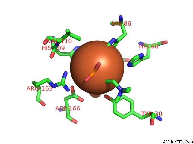
Mono view
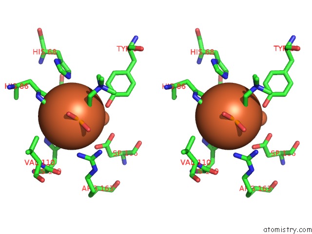
Stereo pair view
|
A full contact list of Iron with other atoms in the Fe binding
site number 10 of X-Ray Crystal Structure of Tmpb, (R)-1-Hydroxy-2- Trimethylaminoethylphosphonate Oxygenase, with (R)-1-Hydroxy-2- Trimethylaminoethylphosphonate within 5.0Å range:
probe
|
atom
|
residue
|
distance (Å)
|
B
|
Occ
|
D:Fe202
b:14.7
occ:1.00
|
O
|
D:HOH313
|
2.0
|
14.6
|
1.0
|
OD1
|
D:ASP65
|
2.2
|
14.2
|
1.0
|
O9
|
D:KVP203
|
2.2
|
18.8
|
1.0
|
NE2
|
D:HIS86
|
2.3
|
19.4
|
1.0
|
NE2
|
D:HIS109
|
2.3
|
15.7
|
1.0
|
O1
|
D:KVP203
|
2.4
|
21.2
|
1.0
|
CG
|
D:ASP65
|
3.2
|
14.2
|
1.0
|
CE1
|
D:HIS86
|
3.3
|
19.8
|
1.0
|
CD2
|
D:HIS86
|
3.3
|
19.4
|
1.0
|
P4
|
D:KVP203
|
3.3
|
20.6
|
1.0
|
CD2
|
D:HIS109
|
3.3
|
15.7
|
1.0
|
C1
|
D:KVP203
|
3.3
|
21.3
|
1.0
|
CE1
|
D:HIS109
|
3.3
|
15.4
|
1.0
|
OD2
|
D:ASP65
|
3.5
|
14.6
|
1.0
|
FE
|
D:FE201
|
3.6
|
15.1
|
1.0
|
OH
|
D:TYR30
|
4.0
|
22.2
|
1.0
|
O10
|
D:KVP203
|
4.1
|
19.9
|
1.0
|
CD2
|
D:HIS64
|
4.2
|
12.9
|
1.0
|
NE2
|
D:HIS68
|
4.2
|
21.7
|
1.0
|
OD2
|
D:ASP166
|
4.3
|
17.5
|
1.0
|
ND1
|
D:HIS86
|
4.4
|
19.9
|
1.0
|
CG
|
D:HIS86
|
4.4
|
19.6
|
1.0
|
O11
|
D:KVP203
|
4.4
|
20.5
|
1.0
|
ND1
|
D:HIS109
|
4.4
|
15.7
|
1.0
|
CG
|
D:HIS109
|
4.4
|
15.9
|
1.0
|
NH1
|
D:ARG163
|
4.5
|
16.3
|
1.0
|
NE2
|
D:HIS64
|
4.5
|
13.0
|
1.0
|
CB
|
D:ASP65
|
4.6
|
13.9
|
1.0
|
C2
|
D:KVP203
|
4.6
|
22.7
|
1.0
|
CG2
|
D:VAL110
|
4.9
|
17.5
|
1.0
|
CD2
|
D:HIS68
|
4.9
|
21.1
|
1.0
|
O
|
D:HIS64
|
4.9
|
11.9
|
1.0
|
CA
|
D:ASP65
|
5.0
|
13.5
|
1.0
|
OD1
|
D:ASP166
|
5.0
|
16.2
|
1.0
|
|
Reference:
L.J.Rajakovich,
M.E.Pandelia,
A.J.Mitchell,
W.C.Chang,
B.Zhang,
A.K.Boal,
C.Krebs,
J.M.Bollinger Jr..
A New Microbial Pathway For Organophosphonate Degradation Catalyzed By Two Previously Misannotated Non-Heme-Iron Oxygenases. Biochemistry V. 58 1627 2019.
ISSN: ISSN 1520-4995
PubMed: 30789718
DOI: 10.1021/ACS.BIOCHEM.9B00044
Page generated: Wed Aug 7 03:47:18 2024
|