Iron »
PDB 6o6m-6op1 »
6o6m »
Iron in PDB 6o6m: The Structure of Egtb (Cabther)
Protein crystallography data
The structure of The Structure of Egtb (Cabther), PDB code: 6o6m
was solved by
S.Irani,
Y.Zhang,
with X-Ray Crystallography technique. A brief refinement statistics is given in the table below:
| Resolution Low / High (Å) | 45.86 / 2.51 |
| Space group | P 1 21 1 |
| Cell size a, b, c (Å), α, β, γ (°) | 85.294, 137.315, 85.391, 90.00, 92.43, 90.00 |
| R / Rfree (%) | 18.5 / 22.6 |
Iron Binding Sites:
The binding sites of Iron atom in the The Structure of Egtb (Cabther)
(pdb code 6o6m). This binding sites where shown within
5.0 Angstroms radius around Iron atom.
In total 4 binding sites of Iron where determined in the The Structure of Egtb (Cabther), PDB code: 6o6m:
Jump to Iron binding site number: 1; 2; 3; 4;
In total 4 binding sites of Iron where determined in the The Structure of Egtb (Cabther), PDB code: 6o6m:
Jump to Iron binding site number: 1; 2; 3; 4;
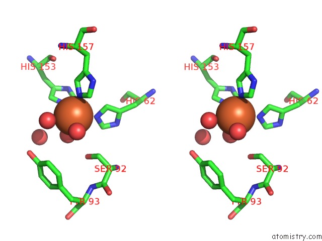
Iron binding site 1 out of 4 in 6o6m
Go back to
Iron binding site 1 out
of 4 in the The Structure of Egtb (Cabther)
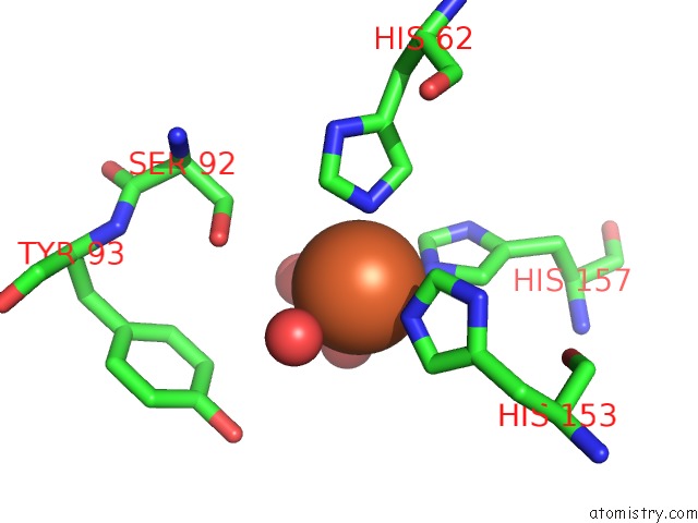
Mono view
Stereo pair view
Mono view
Stereo pair view
A full contact list of Iron with other atoms in the Fe binding
site number 1 of The Structure of Egtb (Cabther) within 5.0Å range:
|
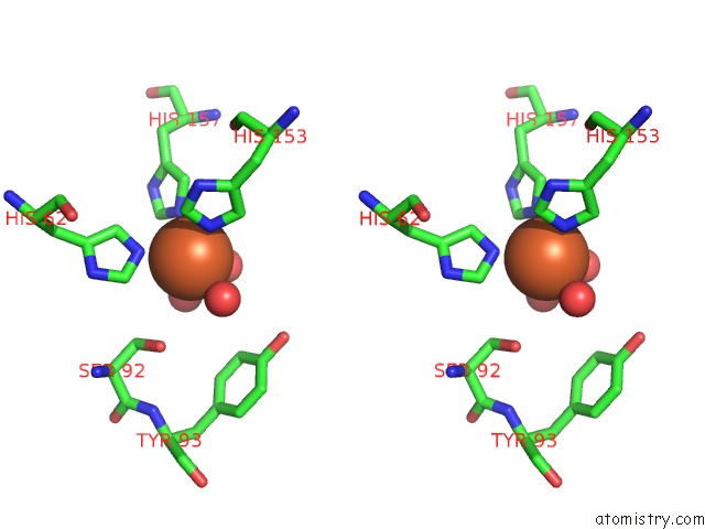
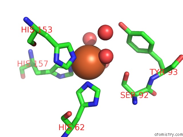
Iron binding site 2 out of 4 in 6o6m
Go back to
Iron binding site 2 out
of 4 in the The Structure of Egtb (Cabther)

Mono view
Stereo pair view
Mono view
Stereo pair view
A full contact list of Iron with other atoms in the Fe binding
site number 2 of The Structure of Egtb (Cabther) within 5.0Å range:
|
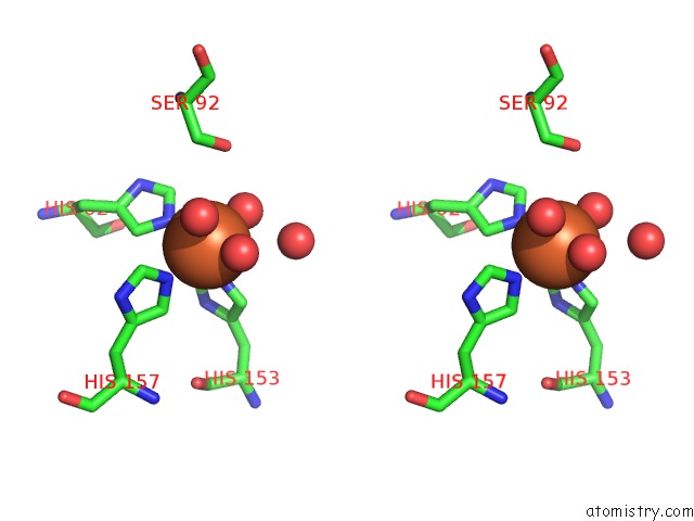
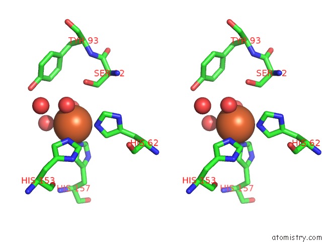
Iron binding site 3 out of 4 in 6o6m
Go back to
Iron binding site 3 out
of 4 in the The Structure of Egtb (Cabther)
Mono view
Stereo pair view
Mono view
Stereo pair view
A full contact list of Iron with other atoms in the Fe binding
site number 3 of The Structure of Egtb (Cabther) within 5.0Å range:
|
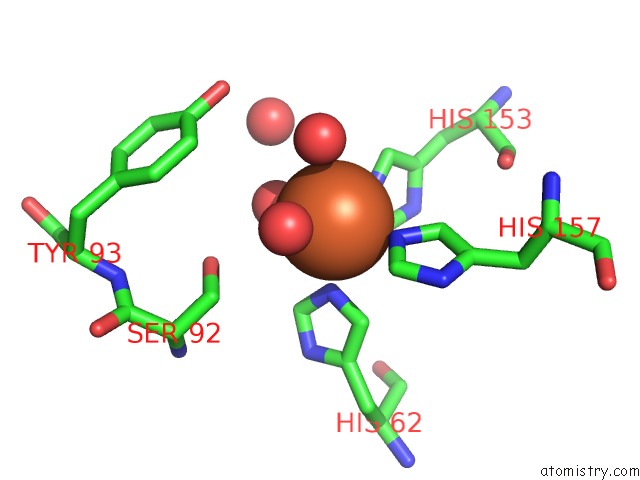
Iron binding site 4 out of 4 in 6o6m
Go back to
Iron binding site 4 out
of 4 in the The Structure of Egtb (Cabther)
Mono view
Stereo pair view
Mono view
Stereo pair view
A full contact list of Iron with other atoms in the Fe binding
site number 4 of The Structure of Egtb (Cabther) within 5.0Å range:
|
Reference:
N.Naowarojna,
S.Irani,
R.Cheng,
W.Hu,
L.Zhang,
X.Li,
J.Chen,
Y.Zhang,
P.Liu.
Crystal Structure of the Ergothioneine Sulfoxide Synthase From Candidatus Chloracidobacterium Thermophilum and Structure-Guided Engineering to Modulate Its Substrate Selectivity Acs Catalysis 2019.
ISSN: ESSN 2155-5435
DOI: 10.1021/ACSCATAL.9B02054
Page generated: Wed Aug 7 04:08:43 2024
ISSN: ESSN 2155-5435
DOI: 10.1021/ACSCATAL.9B02054
Last articles
Zn in 9J0NZn in 9J0O
Zn in 9J0P
Zn in 9FJX
Zn in 9EKB
Zn in 9C0F
Zn in 9CAH
Zn in 9CH0
Zn in 9CH3
Zn in 9CH1