Iron »
PDB 6qgr-6r2o »
6qo7 »
Iron in PDB 6qo7: Crystal Structure of Ribonucleotide Reductase Nrdf From Bacillus Anthracis Aerobically Soaked with Ferrous Ions (Photo-Reduced)
Enzymatic activity of Crystal Structure of Ribonucleotide Reductase Nrdf From Bacillus Anthracis Aerobically Soaked with Ferrous Ions (Photo-Reduced)
All present enzymatic activity of Crystal Structure of Ribonucleotide Reductase Nrdf From Bacillus Anthracis Aerobically Soaked with Ferrous Ions (Photo-Reduced):
1.17.4.1;
1.17.4.1;
Protein crystallography data
The structure of Crystal Structure of Ribonucleotide Reductase Nrdf From Bacillus Anthracis Aerobically Soaked with Ferrous Ions (Photo-Reduced), PDB code: 6qo7
was solved by
K.Grave,
M.Hogbom,
with X-Ray Crystallography technique. A brief refinement statistics is given in the table below:
| Resolution Low / High (Å) | 41.82 / 1.63 |
| Space group | P 1 21 1 |
| Cell size a, b, c (Å), α, β, γ (°) | 57.293, 59.958, 95.360, 90.00, 107.11, 90.00 |
| R / Rfree (%) | 15.9 / 19.2 |
Other elements in 6qo7:
The structure of Crystal Structure of Ribonucleotide Reductase Nrdf From Bacillus Anthracis Aerobically Soaked with Ferrous Ions (Photo-Reduced) also contains other interesting chemical elements:
| Chlorine | (Cl) | 1 atom |
Iron Binding Sites:
The binding sites of Iron atom in the Crystal Structure of Ribonucleotide Reductase Nrdf From Bacillus Anthracis Aerobically Soaked with Ferrous Ions (Photo-Reduced)
(pdb code 6qo7). This binding sites where shown within
5.0 Angstroms radius around Iron atom.
In total 4 binding sites of Iron where determined in the Crystal Structure of Ribonucleotide Reductase Nrdf From Bacillus Anthracis Aerobically Soaked with Ferrous Ions (Photo-Reduced), PDB code: 6qo7:
Jump to Iron binding site number: 1; 2; 3; 4;
In total 4 binding sites of Iron where determined in the Crystal Structure of Ribonucleotide Reductase Nrdf From Bacillus Anthracis Aerobically Soaked with Ferrous Ions (Photo-Reduced), PDB code: 6qo7:
Jump to Iron binding site number: 1; 2; 3; 4;
Iron binding site 1 out of 4 in 6qo7
Go back to
Iron binding site 1 out
of 4 in the Crystal Structure of Ribonucleotide Reductase Nrdf From Bacillus Anthracis Aerobically Soaked with Ferrous Ions (Photo-Reduced)
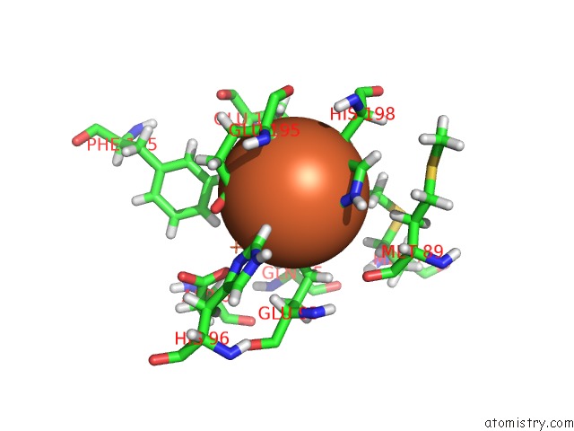
Mono view
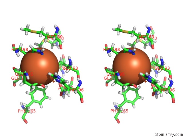
Stereo pair view
Mono view
Stereo pair view
A full contact list of Iron with other atoms in the Fe binding
site number 1 of Crystal Structure of Ribonucleotide Reductase Nrdf From Bacillus Anthracis Aerobically Soaked with Ferrous Ions (Photo-Reduced) within 5.0Å range:
|
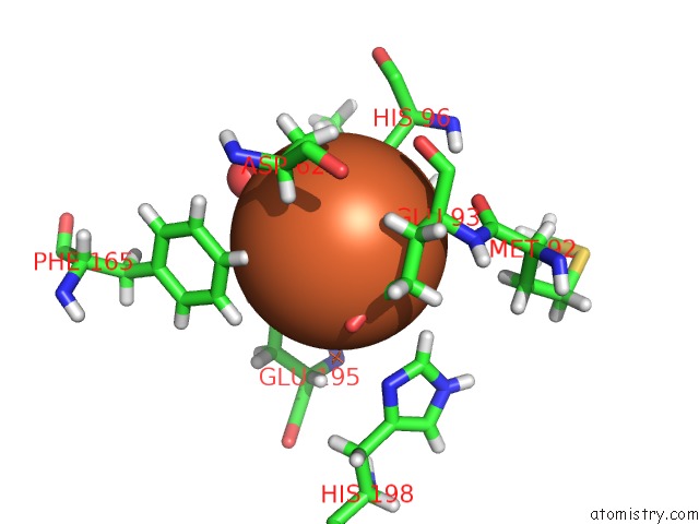
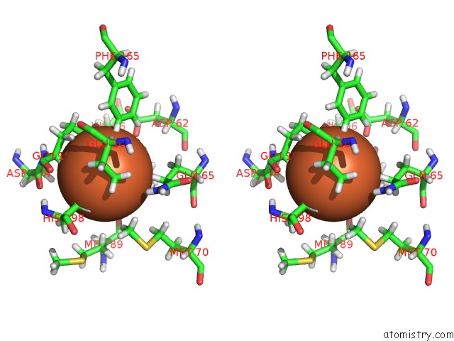
Iron binding site 2 out of 4 in 6qo7
Go back to
Iron binding site 2 out
of 4 in the Crystal Structure of Ribonucleotide Reductase Nrdf From Bacillus Anthracis Aerobically Soaked with Ferrous Ions (Photo-Reduced)

Mono view

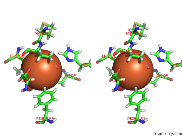
Stereo pair view
Mono view
Stereo pair view
A full contact list of Iron with other atoms in the Fe binding
site number 2 of Crystal Structure of Ribonucleotide Reductase Nrdf From Bacillus Anthracis Aerobically Soaked with Ferrous Ions (Photo-Reduced) within 5.0Å range:
|
Iron binding site 3 out of 4 in 6qo7
Go back to
Iron binding site 3 out
of 4 in the Crystal Structure of Ribonucleotide Reductase Nrdf From Bacillus Anthracis Aerobically Soaked with Ferrous Ions (Photo-Reduced)
Mono view
Stereo pair view
Mono view
Stereo pair view
A full contact list of Iron with other atoms in the Fe binding
site number 3 of Crystal Structure of Ribonucleotide Reductase Nrdf From Bacillus Anthracis Aerobically Soaked with Ferrous Ions (Photo-Reduced) within 5.0Å range:
|
Iron binding site 4 out of 4 in 6qo7
Go back to
Iron binding site 4 out
of 4 in the Crystal Structure of Ribonucleotide Reductase Nrdf From Bacillus Anthracis Aerobically Soaked with Ferrous Ions (Photo-Reduced)

Mono view
Stereo pair view
Mono view
Stereo pair view
A full contact list of Iron with other atoms in the Fe binding
site number 4 of Crystal Structure of Ribonucleotide Reductase Nrdf From Bacillus Anthracis Aerobically Soaked with Ferrous Ions (Photo-Reduced) within 5.0Å range:
|
Reference:
K.Grave,
W.Lambert,
G.Berggren,
J.J.Griese,
M.D.Bennett,
D.T.Logan,
M.Hogbom.
Redox-Induced Structural Changes in the Di-Iron and Di-Manganese Forms of Bacillus Anthracis Ribonucleotide Reductase Subunit Nrdf Suggest A Mechanism For Gating of Radical Access. J.Biol.Inorg.Chem. V. 24 849 2019.
ISSN: ESSN 1432-1327
PubMed: 31410573
DOI: 10.1007/S00775-019-01703-Z
Page generated: Wed Aug 7 08:09:10 2024
ISSN: ESSN 1432-1327
PubMed: 31410573
DOI: 10.1007/S00775-019-01703-Z
Last articles
F in 7QISF in 7QK4
F in 7QK0
F in 7QIR
F in 7QI8
F in 7QC5
F in 7QG3
F in 7QHN
F in 7Q6P
F in 7QBZ