Iron »
PDB 6qgr-6r2o »
6qob »
Iron in PDB 6qob: Crystal Structure of Ribonucleotide Reductase Nrdf From Bacillus Anthracis with Partially Oxidised Di-Iron Metallocofactor
Enzymatic activity of Crystal Structure of Ribonucleotide Reductase Nrdf From Bacillus Anthracis with Partially Oxidised Di-Iron Metallocofactor
All present enzymatic activity of Crystal Structure of Ribonucleotide Reductase Nrdf From Bacillus Anthracis with Partially Oxidised Di-Iron Metallocofactor:
1.17.4.1;
1.17.4.1;
Protein crystallography data
The structure of Crystal Structure of Ribonucleotide Reductase Nrdf From Bacillus Anthracis with Partially Oxidised Di-Iron Metallocofactor, PDB code: 6qob
was solved by
K.Grave,
M.Hogbom,
with X-Ray Crystallography technique. A brief refinement statistics is given in the table below:
| Resolution Low / High (Å) | 45.94 / 1.46 |
| Space group | P 1 21 1 |
| Cell size a, b, c (Å), α, β, γ (°) | 57.137, 60.377, 95.758, 90.00, 106.38, 90.00 |
| R / Rfree (%) | 17 / 19.4 |
Iron Binding Sites:
The binding sites of Iron atom in the Crystal Structure of Ribonucleotide Reductase Nrdf From Bacillus Anthracis with Partially Oxidised Di-Iron Metallocofactor
(pdb code 6qob). This binding sites where shown within
5.0 Angstroms radius around Iron atom.
In total 4 binding sites of Iron where determined in the Crystal Structure of Ribonucleotide Reductase Nrdf From Bacillus Anthracis with Partially Oxidised Di-Iron Metallocofactor, PDB code: 6qob:
Jump to Iron binding site number: 1; 2; 3; 4;
In total 4 binding sites of Iron where determined in the Crystal Structure of Ribonucleotide Reductase Nrdf From Bacillus Anthracis with Partially Oxidised Di-Iron Metallocofactor, PDB code: 6qob:
Jump to Iron binding site number: 1; 2; 3; 4;
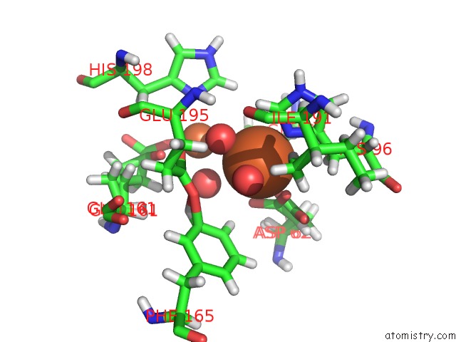
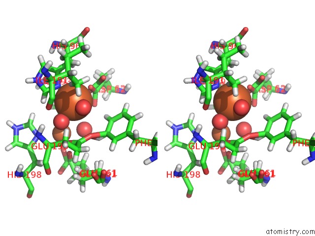
Iron binding site 1 out of 4 in 6qob
Go back to
Iron binding site 1 out
of 4 in the Crystal Structure of Ribonucleotide Reductase Nrdf From Bacillus Anthracis with Partially Oxidised Di-Iron Metallocofactor
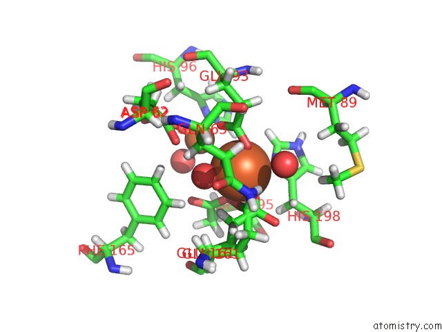
Mono view
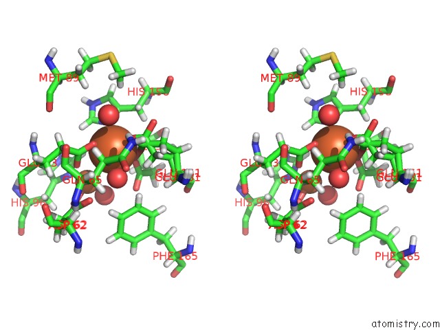
Stereo pair view
Mono view
Stereo pair view
A full contact list of Iron with other atoms in the Fe binding
site number 1 of Crystal Structure of Ribonucleotide Reductase Nrdf From Bacillus Anthracis with Partially Oxidised Di-Iron Metallocofactor within 5.0Å range:
|
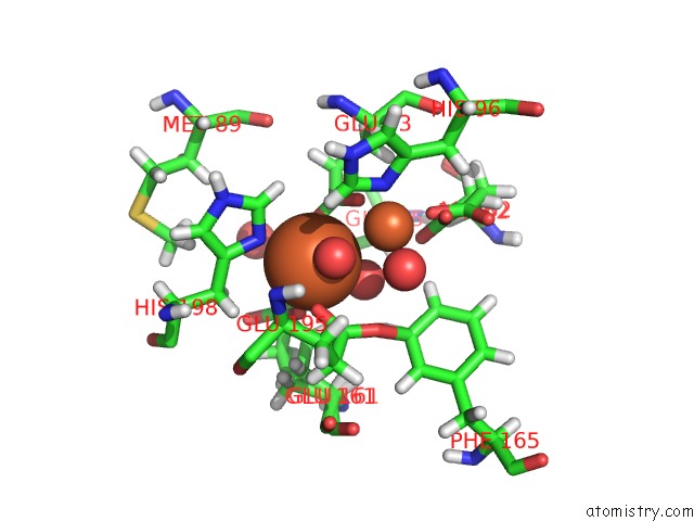
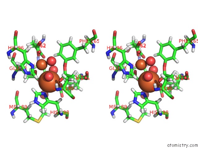
Iron binding site 2 out of 4 in 6qob
Go back to
Iron binding site 2 out
of 4 in the Crystal Structure of Ribonucleotide Reductase Nrdf From Bacillus Anthracis with Partially Oxidised Di-Iron Metallocofactor
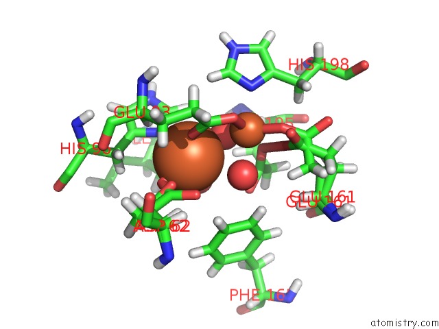
Mono view
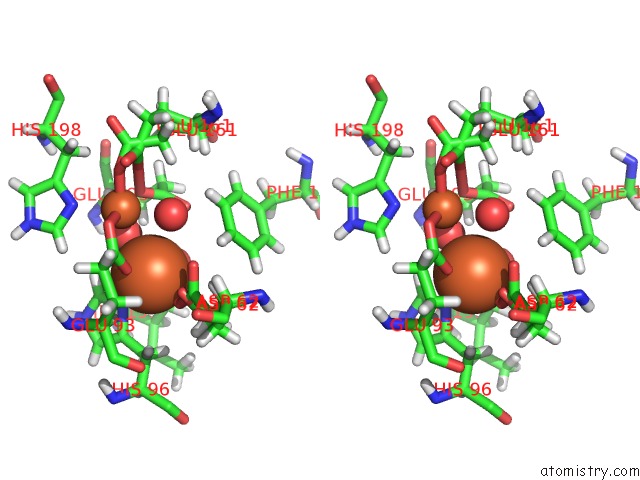
Stereo pair view
Mono view
Stereo pair view
A full contact list of Iron with other atoms in the Fe binding
site number 2 of Crystal Structure of Ribonucleotide Reductase Nrdf From Bacillus Anthracis with Partially Oxidised Di-Iron Metallocofactor within 5.0Å range:
|
Iron binding site 3 out of 4 in 6qob
Go back to
Iron binding site 3 out
of 4 in the Crystal Structure of Ribonucleotide Reductase Nrdf From Bacillus Anthracis with Partially Oxidised Di-Iron Metallocofactor
Mono view
Stereo pair view
Mono view
Stereo pair view
A full contact list of Iron with other atoms in the Fe binding
site number 3 of Crystal Structure of Ribonucleotide Reductase Nrdf From Bacillus Anthracis with Partially Oxidised Di-Iron Metallocofactor within 5.0Å range:
|
Iron binding site 4 out of 4 in 6qob
Go back to
Iron binding site 4 out
of 4 in the Crystal Structure of Ribonucleotide Reductase Nrdf From Bacillus Anthracis with Partially Oxidised Di-Iron Metallocofactor
Mono view
Stereo pair view
Mono view
Stereo pair view
A full contact list of Iron with other atoms in the Fe binding
site number 4 of Crystal Structure of Ribonucleotide Reductase Nrdf From Bacillus Anthracis with Partially Oxidised Di-Iron Metallocofactor within 5.0Å range:
|
Reference:
K.Grave,
W.Lambert,
G.Berggren,
J.J.Griese,
M.D.Bennett,
D.T.Logan,
M.Hogbom.
Redox-Induced Structural Changes in the Di-Iron and Di-Manganese Forms of Bacillus Anthracis Ribonucleotide Reductase Subunit Nrdf Suggest A Mechanism For Gating of Radical Access. J.Biol.Inorg.Chem. V. 24 849 2019.
ISSN: ESSN 1432-1327
PubMed: 31410573
DOI: 10.1007/S00775-019-01703-Z
Page generated: Wed Aug 7 08:11:31 2024
ISSN: ESSN 1432-1327
PubMed: 31410573
DOI: 10.1007/S00775-019-01703-Z
Last articles
Zn in 9MJ5Zn in 9HNW
Zn in 9G0L
Zn in 9FNE
Zn in 9DZN
Zn in 9E0I
Zn in 9D32
Zn in 9DAK
Zn in 8ZXC
Zn in 8ZUF