Iron »
PDB 6r2q-6rr1 »
6ro8 »
Iron in PDB 6ro8: The Crystal Structure of Acinetobacter Radioresistens CYP116B5 Heme Domain
Protein crystallography data
The structure of The Crystal Structure of Acinetobacter Radioresistens CYP116B5 Heme Domain, PDB code: 6ro8
was solved by
A.Ciaramella,
G.Catucci,
G.Gilardi,
G.Di Nardo,
with X-Ray Crystallography technique. A brief refinement statistics is given in the table below:
| Resolution Low / High (Å) | 91.33 / 2.60 |
| Space group | P 41 21 2 |
| Cell size a, b, c (Å), α, β, γ (°) | 109.900, 109.900, 164.200, 90.00, 90.00, 90.00 |
| R / Rfree (%) | 20 / 24 |
Iron Binding Sites:
The binding sites of Iron atom in the The Crystal Structure of Acinetobacter Radioresistens CYP116B5 Heme Domain
(pdb code 6ro8). This binding sites where shown within
5.0 Angstroms radius around Iron atom.
In total only one binding site of Iron was determined in the The Crystal Structure of Acinetobacter Radioresistens CYP116B5 Heme Domain, PDB code: 6ro8:
In total only one binding site of Iron was determined in the The Crystal Structure of Acinetobacter Radioresistens CYP116B5 Heme Domain, PDB code: 6ro8:
Iron binding site 1 out of 1 in 6ro8
Go back to
Iron binding site 1 out
of 1 in the The Crystal Structure of Acinetobacter Radioresistens CYP116B5 Heme Domain
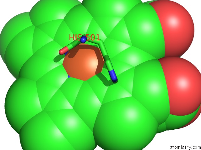
Mono view
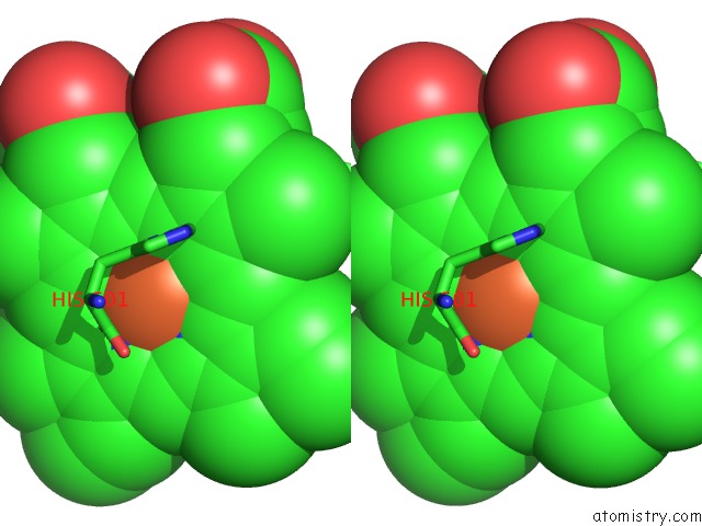
Stereo pair view
Mono view
Stereo pair view
A full contact list of Iron with other atoms in the Fe binding
site number 1 of The Crystal Structure of Acinetobacter Radioresistens CYP116B5 Heme Domain within 5.0Å range:
|
Reference:
A.Ciaramella,
G.Catucci,
G.Gilardi,
G.Di Nardo.
Crystal Structure of Bacterial CYP116B5 Heme Domain: New Insights on Class VII P450S Structural Flexibility and Peroxygenase Activity. Int.J.Biol.Macromol. V. 140 577 2019.
ISSN: ISSN 0141-8130
PubMed: 31430491
DOI: 10.1016/J.IJBIOMAC.2019.08.141
Page generated: Wed Aug 7 08:51:25 2024
ISSN: ISSN 0141-8130
PubMed: 31430491
DOI: 10.1016/J.IJBIOMAC.2019.08.141
Last articles
Fe in 2YXOFe in 2YRS
Fe in 2YXC
Fe in 2YNM
Fe in 2YVJ
Fe in 2YP1
Fe in 2YU2
Fe in 2YU1
Fe in 2YQB
Fe in 2YOO