Iron »
PDB 6sor-6tjv »
6soz »
Iron in PDB 6soz: Glycosylated Trypanosoma Brucei Transferrin Receptor in Complex with Human Transferrin
Protein crystallography data
The structure of Glycosylated Trypanosoma Brucei Transferrin Receptor in Complex with Human Transferrin, PDB code: 6soz
was solved by
C.Trevor,
M.Carrington,
M.K.Higgins,
with X-Ray Crystallography technique. A brief refinement statistics is given in the table below:
| Resolution Low / High (Å) | 39.59 / 3.42 |
| Space group | C 1 2 1 |
| Cell size a, b, c (Å), α, β, γ (°) | 128.180, 117.870, 134.550, 90.00, 111.45, 90.00 |
| R / Rfree (%) | 21.1 / 24.1 |
Iron Binding Sites:
The binding sites of Iron atom in the Glycosylated Trypanosoma Brucei Transferrin Receptor in Complex with Human Transferrin
(pdb code 6soz). This binding sites where shown within
5.0 Angstroms radius around Iron atom.
In total only one binding site of Iron was determined in the Glycosylated Trypanosoma Brucei Transferrin Receptor in Complex with Human Transferrin, PDB code: 6soz:
In total only one binding site of Iron was determined in the Glycosylated Trypanosoma Brucei Transferrin Receptor in Complex with Human Transferrin, PDB code: 6soz:
Iron binding site 1 out of 1 in 6soz
Go back to
Iron binding site 1 out
of 1 in the Glycosylated Trypanosoma Brucei Transferrin Receptor in Complex with Human Transferrin
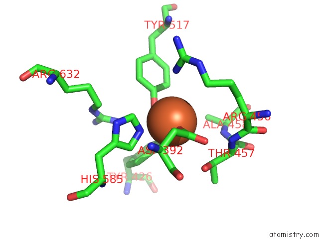
Mono view
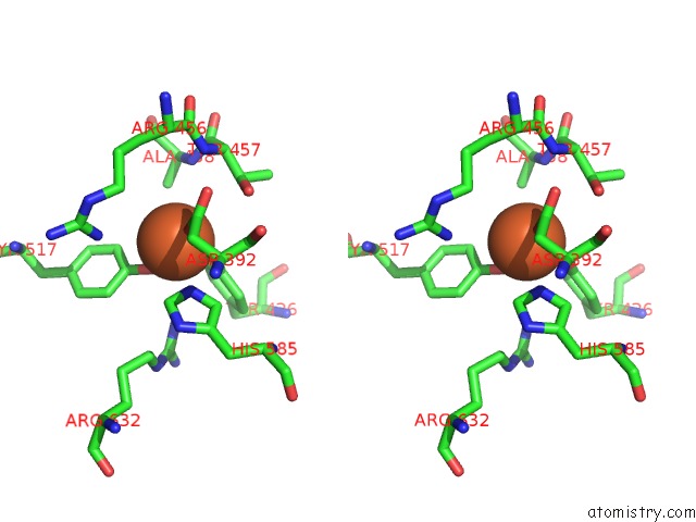
Stereo pair view
Mono view
Stereo pair view
A full contact list of Iron with other atoms in the Fe binding
site number 1 of Glycosylated Trypanosoma Brucei Transferrin Receptor in Complex with Human Transferrin within 5.0Å range:
|
Reference:
C.E.Trevor,
A.L.Gonzalez-Munoz,
O.J.S.Macleod,
P.G.Woodcock,
S.Rust,
T.J.Vaughan,
E.F.Garman,
R.Minter,
M.Carrington,
M.K.Higgins.
Structure of the Trypanosome Transferrin Receptor Reveals Mechanisms of Ligand Recognition and Immune Evasion. Nat Microbiol V. 4 2074 2019.
ISSN: ESSN 2058-5276
PubMed: 31636418
DOI: 10.1038/S41564-019-0589-0
Page generated: Wed Aug 7 10:34:32 2024
ISSN: ESSN 2058-5276
PubMed: 31636418
DOI: 10.1038/S41564-019-0589-0
Last articles
F in 4MS5F in 4MOS
F in 4MOR
F in 4MNE
F in 4MOP
F in 4MOQ
F in 4MOO
F in 4MOM
F in 4MOL
F in 4MOJ