Iron »
PDB 6sor-6tjv »
6tb5 »
Iron in PDB 6tb5: The Crystal Structure of the DPS2 From Deinococcus Radiodurans to 1.83A Resolution (Sequentially Soaked in CACL2 [5MM] For 20 Min, Then in Ammonium Iron(II) Sulfate [10MM] For 2H).
Protein crystallography data
The structure of The Crystal Structure of the DPS2 From Deinococcus Radiodurans to 1.83A Resolution (Sequentially Soaked in CACL2 [5MM] For 20 Min, Then in Ammonium Iron(II) Sulfate [10MM] For 2H)., PDB code: 6tb5
was solved by
M.G.Cuypers,
S.Mcsweeney,
C.V.Romao,
E.P.Mitchell,
with X-Ray Crystallography technique. A brief refinement statistics is given in the table below:
| Resolution Low / High (Å) | 28.09 / 1.83 |
| Space group | P 2 3 |
| Cell size a, b, c (Å), α, β, γ (°) | 88.830, 88.830, 88.830, 90.00, 90.00, 90.00 |
| R / Rfree (%) | 17.3 / 21.2 |
Other elements in 6tb5:
The structure of The Crystal Structure of the DPS2 From Deinococcus Radiodurans to 1.83A Resolution (Sequentially Soaked in CACL2 [5MM] For 20 Min, Then in Ammonium Iron(II) Sulfate [10MM] For 2H). also contains other interesting chemical elements:
| Calcium | (Ca) | 2 atoms |
Iron Binding Sites:
The binding sites of Iron atom in the The Crystal Structure of the DPS2 From Deinococcus Radiodurans to 1.83A Resolution (Sequentially Soaked in CACL2 [5MM] For 20 Min, Then in Ammonium Iron(II) Sulfate [10MM] For 2H).
(pdb code 6tb5). This binding sites where shown within
5.0 Angstroms radius around Iron atom.
In total 4 binding sites of Iron where determined in the The Crystal Structure of the DPS2 From Deinococcus Radiodurans to 1.83A Resolution (Sequentially Soaked in CACL2 [5MM] For 20 Min, Then in Ammonium Iron(II) Sulfate [10MM] For 2H)., PDB code: 6tb5:
Jump to Iron binding site number: 1; 2; 3; 4;
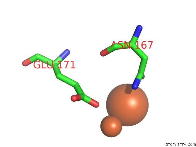
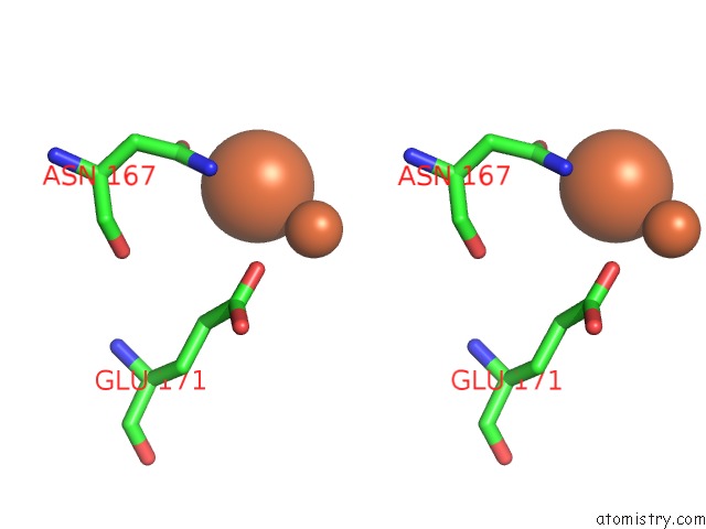
Iron binding site 1 out of 4 in 6tb5
Go back to
Iron binding site 1 out
of 4 in the The Crystal Structure of the DPS2 From Deinococcus Radiodurans to 1.83A Resolution (Sequentially Soaked in CACL2 [5MM] For 20 Min, Then in Ammonium Iron(II) Sulfate [10MM] For 2H).
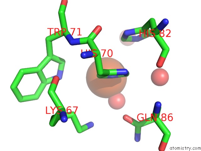
Mono view
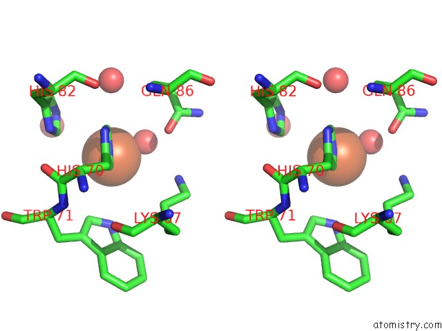
Stereo pair view
Mono view
Stereo pair view
A full contact list of Iron with other atoms in the Fe binding
site number 1 of The Crystal Structure of the DPS2 From Deinococcus Radiodurans to 1.83A Resolution (Sequentially Soaked in CACL2 [5MM] For 20 Min, Then in Ammonium Iron(II) Sulfate [10MM] For 2H). within 5.0Å range:
|
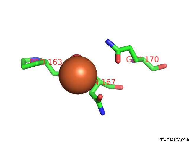
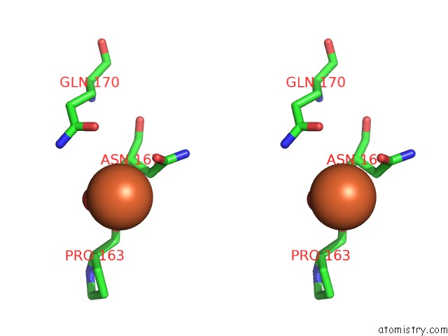
Iron binding site 2 out of 4 in 6tb5
Go back to
Iron binding site 2 out
of 4 in the The Crystal Structure of the DPS2 From Deinococcus Radiodurans to 1.83A Resolution (Sequentially Soaked in CACL2 [5MM] For 20 Min, Then in Ammonium Iron(II) Sulfate [10MM] For 2H).
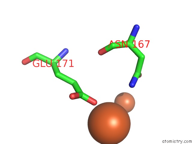
Mono view
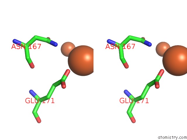
Stereo pair view
Mono view
Stereo pair view
A full contact list of Iron with other atoms in the Fe binding
site number 2 of The Crystal Structure of the DPS2 From Deinococcus Radiodurans to 1.83A Resolution (Sequentially Soaked in CACL2 [5MM] For 20 Min, Then in Ammonium Iron(II) Sulfate [10MM] For 2H). within 5.0Å range:
|
Iron binding site 3 out of 4 in 6tb5
Go back to
Iron binding site 3 out
of 4 in the The Crystal Structure of the DPS2 From Deinococcus Radiodurans to 1.83A Resolution (Sequentially Soaked in CACL2 [5MM] For 20 Min, Then in Ammonium Iron(II) Sulfate [10MM] For 2H).
Mono view
Stereo pair view
Mono view
Stereo pair view
A full contact list of Iron with other atoms in the Fe binding
site number 3 of The Crystal Structure of the DPS2 From Deinococcus Radiodurans to 1.83A Resolution (Sequentially Soaked in CACL2 [5MM] For 20 Min, Then in Ammonium Iron(II) Sulfate [10MM] For 2H). within 5.0Å range:
|
Iron binding site 4 out of 4 in 6tb5
Go back to
Iron binding site 4 out
of 4 in the The Crystal Structure of the DPS2 From Deinococcus Radiodurans to 1.83A Resolution (Sequentially Soaked in CACL2 [5MM] For 20 Min, Then in Ammonium Iron(II) Sulfate [10MM] For 2H).
Mono view
Stereo pair view
Mono view
Stereo pair view
A full contact list of Iron with other atoms in the Fe binding
site number 4 of The Crystal Structure of the DPS2 From Deinococcus Radiodurans to 1.83A Resolution (Sequentially Soaked in CACL2 [5MM] For 20 Min, Then in Ammonium Iron(II) Sulfate [10MM] For 2H). within 5.0Å range:
|
Reference:
M.G.Cuypers,
S.Mcsweeney,
C.V.Romao,
E.P.Mitchell.
The Crystal Structure of the DPS2 From Deinococcus Radiodurans to 1.83A Resolution (Sequentially Soaked in CACL2 [5MM] For 20 Min, Then in Ammonium Iron(II) Sulfate [10MM] For 2H). To Be Published.
Page generated: Wed Aug 7 10:41:04 2024
Last articles
Fe in 2YXOFe in 2YRS
Fe in 2YXC
Fe in 2YNM
Fe in 2YVJ
Fe in 2YP1
Fe in 2YU2
Fe in 2YU1
Fe in 2YQB
Fe in 2YOO