Iron »
PDB 6sor-6tjv »
6tga »
Iron in PDB 6tga: Cryo-Em Structure of As Isolated Form of Nad+-Dependent Formate Dehydrogenase From Rhodobacter Capsulatus
Enzymatic activity of Cryo-Em Structure of As Isolated Form of Nad+-Dependent Formate Dehydrogenase From Rhodobacter Capsulatus
All present enzymatic activity of Cryo-Em Structure of As Isolated Form of Nad+-Dependent Formate Dehydrogenase From Rhodobacter Capsulatus:
1.2.1.2;
1.2.1.2;
Other elements in 6tga:
The structure of Cryo-Em Structure of As Isolated Form of Nad+-Dependent Formate Dehydrogenase From Rhodobacter Capsulatus also contains other interesting chemical elements:
| Molybdenum | (Mo) | 2 atoms |
Iron Binding Sites:
Pages:
>>> Page 1 <<< Page 2, Binding sites: 11 - 20; Page 3, Binding sites: 21 - 30; Page 4, Binding sites: 31 - 40; Page 5, Binding sites: 41 - 48;Binding sites:
The binding sites of Iron atom in the Cryo-Em Structure of As Isolated Form of Nad+-Dependent Formate Dehydrogenase From Rhodobacter Capsulatus (pdb code 6tga). This binding sites where shown within 5.0 Angstroms radius around Iron atom.In total 48 binding sites of Iron where determined in the Cryo-Em Structure of As Isolated Form of Nad+-Dependent Formate Dehydrogenase From Rhodobacter Capsulatus, PDB code: 6tga:
Jump to Iron binding site number: 1; 2; 3; 4; 5; 6; 7; 8; 9; 10;
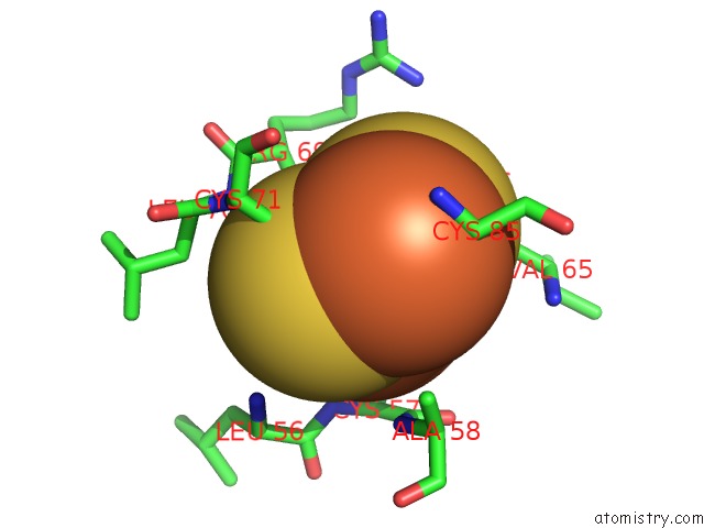
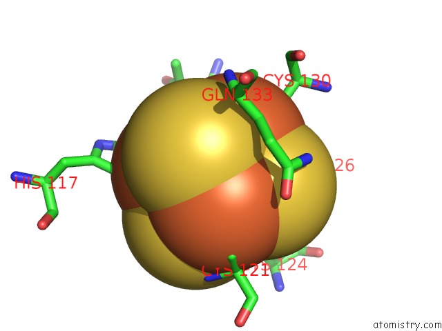
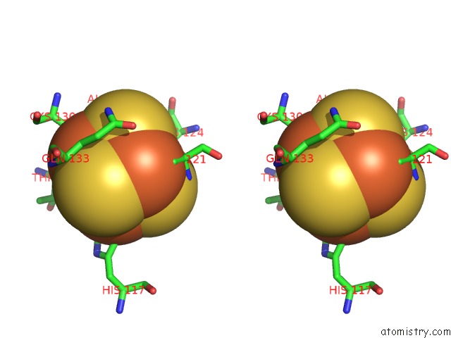
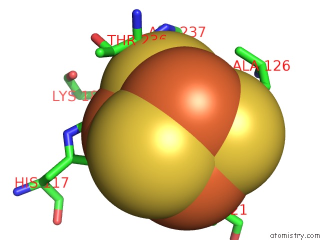
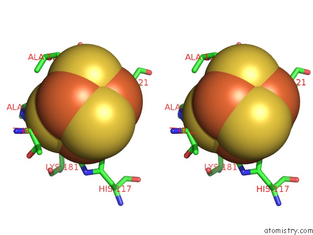
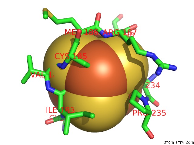
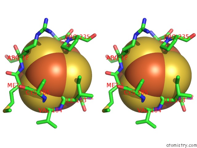
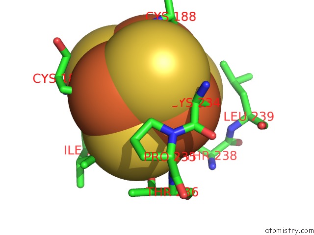
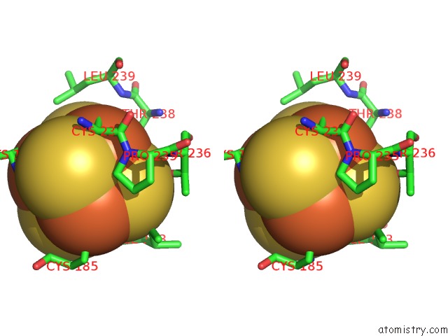
Iron binding site 1 out of 48 in 6tga
Go back to
Iron binding site 1 out
of 48 in the Cryo-Em Structure of As Isolated Form of Nad+-Dependent Formate Dehydrogenase From Rhodobacter Capsulatus
Mono view
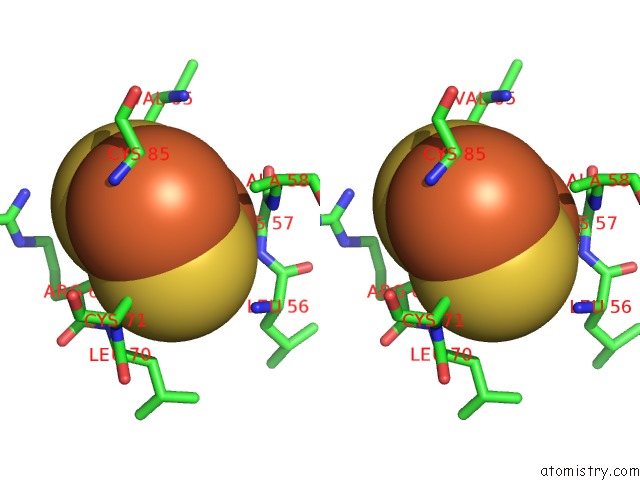
Stereo pair view
Mono view
Stereo pair view
A full contact list of Iron with other atoms in the Fe binding
site number 1 of Cryo-Em Structure of As Isolated Form of Nad+-Dependent Formate Dehydrogenase From Rhodobacter Capsulatus within 5.0Å range:
|
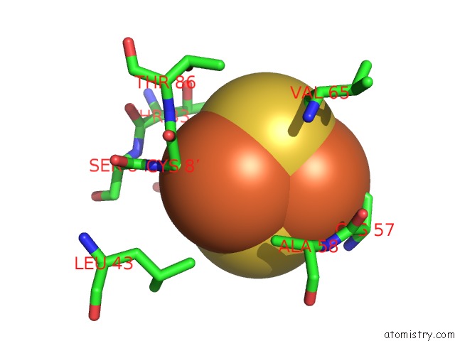
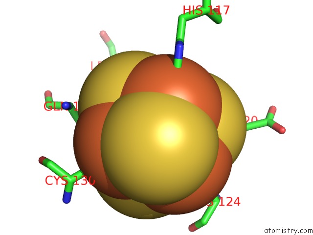
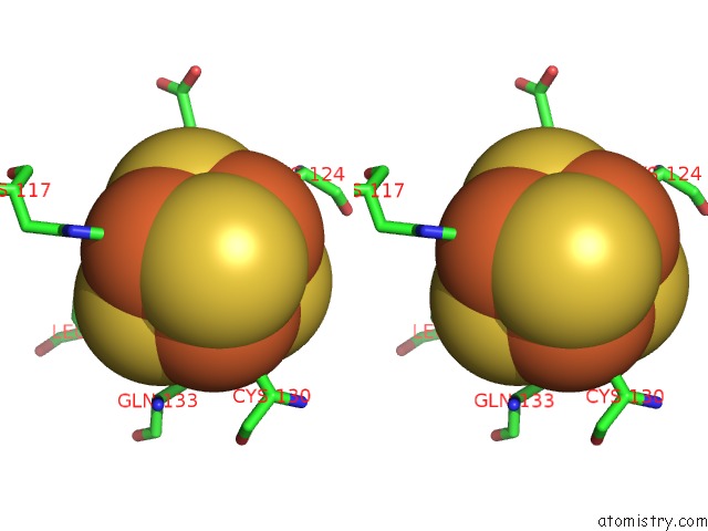
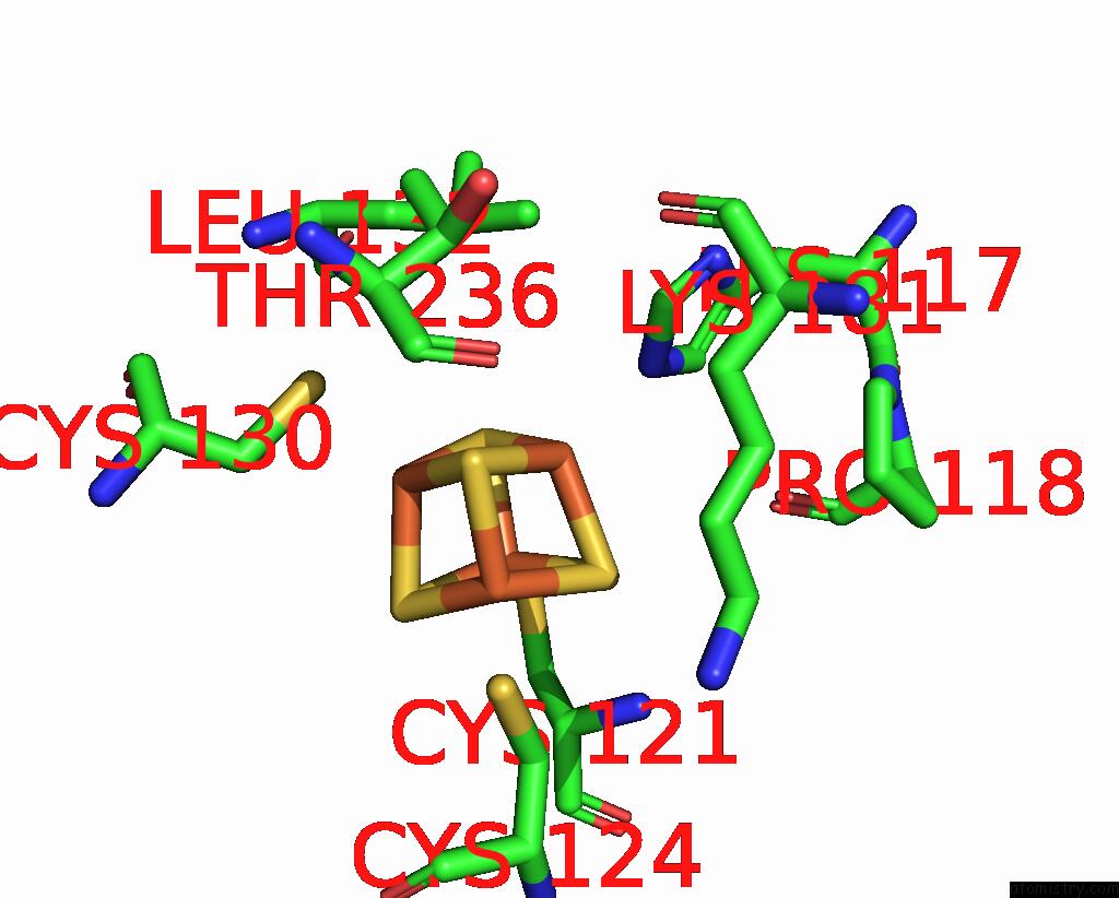
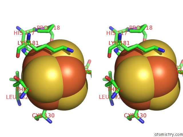
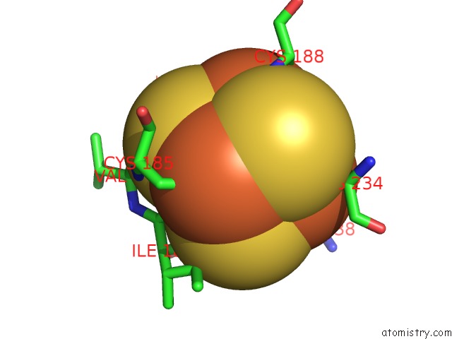
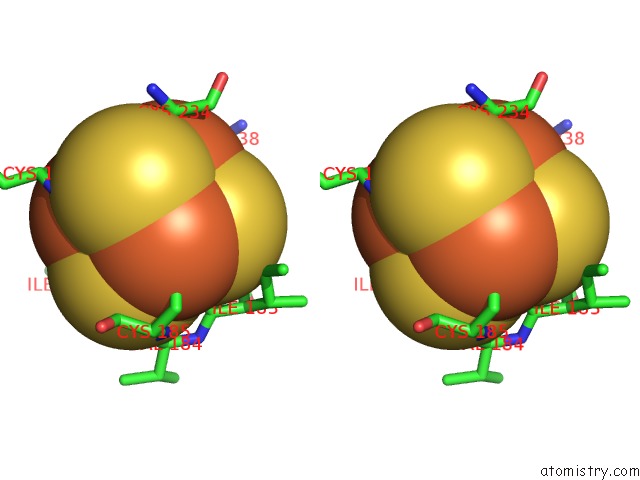
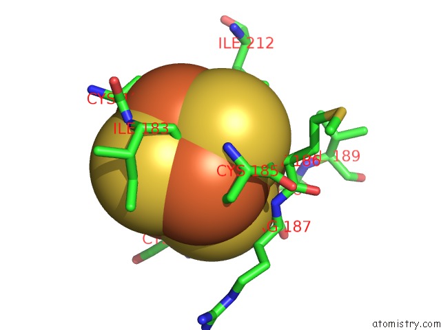
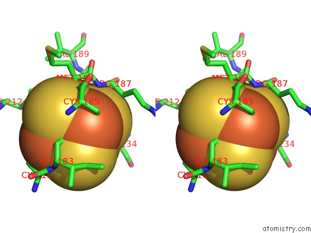
Iron binding site 2 out of 48 in 6tga
Go back to
Iron binding site 2 out
of 48 in the Cryo-Em Structure of As Isolated Form of Nad+-Dependent Formate Dehydrogenase From Rhodobacter Capsulatus
Mono view
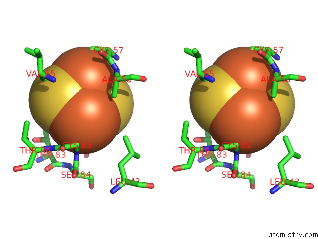
Stereo pair view
Mono view
Stereo pair view
A full contact list of Iron with other atoms in the Fe binding
site number 2 of Cryo-Em Structure of As Isolated Form of Nad+-Dependent Formate Dehydrogenase From Rhodobacter Capsulatus within 5.0Å range:
|
Iron binding site 3 out of 48 in 6tga
Go back to
Iron binding site 3 out
of 48 in the Cryo-Em Structure of As Isolated Form of Nad+-Dependent Formate Dehydrogenase From Rhodobacter Capsulatus
Mono view
Stereo pair view
Mono view
Stereo pair view
A full contact list of Iron with other atoms in the Fe binding
site number 3 of Cryo-Em Structure of As Isolated Form of Nad+-Dependent Formate Dehydrogenase From Rhodobacter Capsulatus within 5.0Å range:
|
Iron binding site 4 out of 48 in 6tga
Go back to
Iron binding site 4 out
of 48 in the Cryo-Em Structure of As Isolated Form of Nad+-Dependent Formate Dehydrogenase From Rhodobacter Capsulatus
Mono view
Stereo pair view
Mono view
Stereo pair view
A full contact list of Iron with other atoms in the Fe binding
site number 4 of Cryo-Em Structure of As Isolated Form of Nad+-Dependent Formate Dehydrogenase From Rhodobacter Capsulatus within 5.0Å range:
|
Iron binding site 5 out of 48 in 6tga
Go back to
Iron binding site 5 out
of 48 in the Cryo-Em Structure of As Isolated Form of Nad+-Dependent Formate Dehydrogenase From Rhodobacter Capsulatus
Mono view
Stereo pair view
Mono view
Stereo pair view
A full contact list of Iron with other atoms in the Fe binding
site number 5 of Cryo-Em Structure of As Isolated Form of Nad+-Dependent Formate Dehydrogenase From Rhodobacter Capsulatus within 5.0Å range:
|
Iron binding site 6 out of 48 in 6tga
Go back to
Iron binding site 6 out
of 48 in the Cryo-Em Structure of As Isolated Form of Nad+-Dependent Formate Dehydrogenase From Rhodobacter Capsulatus
Mono view
Stereo pair view
Mono view
Stereo pair view
A full contact list of Iron with other atoms in the Fe binding
site number 6 of Cryo-Em Structure of As Isolated Form of Nad+-Dependent Formate Dehydrogenase From Rhodobacter Capsulatus within 5.0Å range:
|
Iron binding site 7 out of 48 in 6tga
Go back to
Iron binding site 7 out
of 48 in the Cryo-Em Structure of As Isolated Form of Nad+-Dependent Formate Dehydrogenase From Rhodobacter Capsulatus
Mono view
Stereo pair view
Mono view
Stereo pair view
A full contact list of Iron with other atoms in the Fe binding
site number 7 of Cryo-Em Structure of As Isolated Form of Nad+-Dependent Formate Dehydrogenase From Rhodobacter Capsulatus within 5.0Å range:
|
Iron binding site 8 out of 48 in 6tga
Go back to
Iron binding site 8 out
of 48 in the Cryo-Em Structure of As Isolated Form of Nad+-Dependent Formate Dehydrogenase From Rhodobacter Capsulatus
Mono view
Stereo pair view
Mono view
Stereo pair view
A full contact list of Iron with other atoms in the Fe binding
site number 8 of Cryo-Em Structure of As Isolated Form of Nad+-Dependent Formate Dehydrogenase From Rhodobacter Capsulatus within 5.0Å range:
|
Iron binding site 9 out of 48 in 6tga
Go back to
Iron binding site 9 out
of 48 in the Cryo-Em Structure of As Isolated Form of Nad+-Dependent Formate Dehydrogenase From Rhodobacter Capsulatus
Mono view
Stereo pair view
Mono view
Stereo pair view
A full contact list of Iron with other atoms in the Fe binding
site number 9 of Cryo-Em Structure of As Isolated Form of Nad+-Dependent Formate Dehydrogenase From Rhodobacter Capsulatus within 5.0Å range:
|
Iron binding site 10 out of 48 in 6tga
Go back to
Iron binding site 10 out
of 48 in the Cryo-Em Structure of As Isolated Form of Nad+-Dependent Formate Dehydrogenase From Rhodobacter Capsulatus
Mono view
Stereo pair view
Mono view
Stereo pair view
A full contact list of Iron with other atoms in the Fe binding
site number 10 of Cryo-Em Structure of As Isolated Form of Nad+-Dependent Formate Dehydrogenase From Rhodobacter Capsulatus within 5.0Å range:
|
Reference:
P.Wendler,
C.Radon,
G.Mittelstaedt,
B.R.Duffus,
J.Buerger,
T.Mielke,
S.Leimkuehler.
Cryo-Em Structures Reveal Intricate Fe-S Cluster Arrangement and Charging in Rhodobacter Capsulatus Formate Dehydrogenase Nat Commun 2020.
ISSN: ESSN 2041-1723
DOI: 10.1038/S41467-020-15614-0
Page generated: Wed Aug 7 10:52:46 2024
ISSN: ESSN 2041-1723
DOI: 10.1038/S41467-020-15614-0
Last articles
Zn in 9MJ5Zn in 9HNW
Zn in 9G0L
Zn in 9FNE
Zn in 9DZN
Zn in 9E0I
Zn in 9D32
Zn in 9DAK
Zn in 8ZXC
Zn in 8ZUF