Iron »
PDB 6u97-6uw2 »
6ukl »
Iron in PDB 6ukl: Crystal Structure of A DIB2-Split Protein
Protein crystallography data
The structure of Crystal Structure of A DIB2-Split Protein, PDB code: 6ukl
was solved by
N.G.Bozhanova,
J.Meiler,
with X-Ray Crystallography technique. A brief refinement statistics is given in the table below:
| Resolution Low / High (Å) | 72.10 / 2.02 |
| Space group | P 32 2 1 |
| Cell size a, b, c (Å), α, β, γ (°) | 68.241, 68.241, 216.292, 90.00, 90.00, 120.00 |
| R / Rfree (%) | 20.5 / 24.8 |
Iron Binding Sites:
The binding sites of Iron atom in the Crystal Structure of A DIB2-Split Protein
(pdb code 6ukl). This binding sites where shown within
5.0 Angstroms radius around Iron atom.
In total only one binding site of Iron was determined in the Crystal Structure of A DIB2-Split Protein, PDB code: 6ukl:
In total only one binding site of Iron was determined in the Crystal Structure of A DIB2-Split Protein, PDB code: 6ukl:
Iron binding site 1 out of 1 in 6ukl
Go back to
Iron binding site 1 out
of 1 in the Crystal Structure of A DIB2-Split Protein
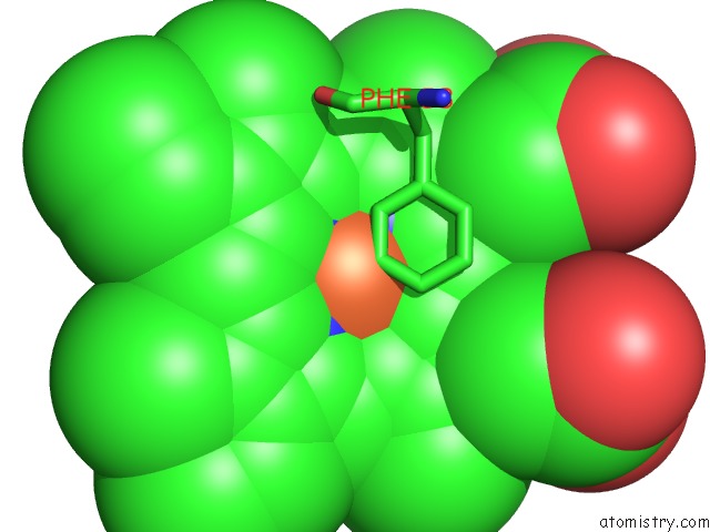
Mono view
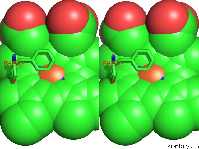
Stereo pair view
Mono view
Stereo pair view
A full contact list of Iron with other atoms in the Fe binding
site number 1 of Crystal Structure of A DIB2-Split Protein within 5.0Å range:
|
Reference:
N.G.Bozhanova,
A.S.Gavrikov,
A.S.Mishin,
J.Meiler.
Dib-Splits: Nature-Guided Design of A Novel Fluorescent Labeling Split System. Sci Rep V. 10 11049 2020.
ISSN: ESSN 2045-2322
PubMed: 32632329
DOI: 10.1038/S41598-020-67095-2
Page generated: Wed Aug 7 12:18:37 2024
ISSN: ESSN 2045-2322
PubMed: 32632329
DOI: 10.1038/S41598-020-67095-2
Last articles
Zn in 9MJ5Zn in 9HNW
Zn in 9G0L
Zn in 9FNE
Zn in 9DZN
Zn in 9E0I
Zn in 9D32
Zn in 9DAK
Zn in 8ZXC
Zn in 8ZUF