Iron »
PDB 6u97-6uw2 »
6upi »
Iron in PDB 6upi: Crystal Structure of Mycobacterium Tuberculosis CYP121 Bound with A Hydroxylated Intermediate of Cyf-4-Ome
Enzymatic activity of Crystal Structure of Mycobacterium Tuberculosis CYP121 Bound with A Hydroxylated Intermediate of Cyf-4-Ome
All present enzymatic activity of Crystal Structure of Mycobacterium Tuberculosis CYP121 Bound with A Hydroxylated Intermediate of Cyf-4-Ome:
1.14.19.70;
1.14.19.70;
Protein crystallography data
The structure of Crystal Structure of Mycobacterium Tuberculosis CYP121 Bound with A Hydroxylated Intermediate of Cyf-4-Ome, PDB code: 6upi
was solved by
R.C.D.Nguyen,
Y.Yang,
A.Liu,
with X-Ray Crystallography technique. A brief refinement statistics is given in the table below:
| Resolution Low / High (Å) | 46.84 / 1.81 |
| Space group | P 65 2 2 |
| Cell size a, b, c (Å), α, β, γ (°) | 77.370, 77.370, 261.973, 90.00, 90.00, 120.00 |
| R / Rfree (%) | 18.5 / 22.5 |
Iron Binding Sites:
The binding sites of Iron atom in the Crystal Structure of Mycobacterium Tuberculosis CYP121 Bound with A Hydroxylated Intermediate of Cyf-4-Ome
(pdb code 6upi). This binding sites where shown within
5.0 Angstroms radius around Iron atom.
In total only one binding site of Iron was determined in the Crystal Structure of Mycobacterium Tuberculosis CYP121 Bound with A Hydroxylated Intermediate of Cyf-4-Ome, PDB code: 6upi:
In total only one binding site of Iron was determined in the Crystal Structure of Mycobacterium Tuberculosis CYP121 Bound with A Hydroxylated Intermediate of Cyf-4-Ome, PDB code: 6upi:
Iron binding site 1 out of 1 in 6upi
Go back to
Iron binding site 1 out
of 1 in the Crystal Structure of Mycobacterium Tuberculosis CYP121 Bound with A Hydroxylated Intermediate of Cyf-4-Ome
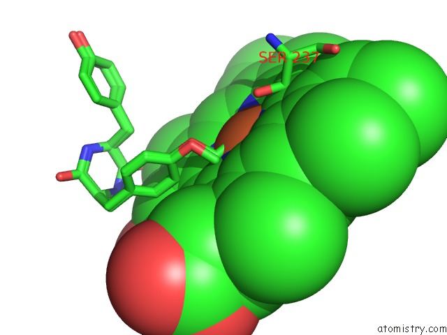
Mono view
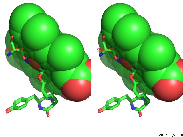
Stereo pair view
Mono view
Stereo pair view
A full contact list of Iron with other atoms in the Fe binding
site number 1 of Crystal Structure of Mycobacterium Tuberculosis CYP121 Bound with A Hydroxylated Intermediate of Cyf-4-Ome within 5.0Å range:
|
Reference:
R.C.Nguyen,
Y.Yang,
I.Davis,
A.Liu.
Substrate-Assisted Hydroxylation and O-Demethylation in the Peroxidase-Like Cytochrome P450 Enzyme CYP121 Acs Catalysis V. 10 1628 2020.
ISSN: ESSN 2155-5435
DOI: 10.1021/ACSCATAL.9B04596
Page generated: Wed Aug 7 12:24:30 2024
ISSN: ESSN 2155-5435
DOI: 10.1021/ACSCATAL.9B04596
Last articles
Zn in 9MJ5Zn in 9HNW
Zn in 9G0L
Zn in 9FNE
Zn in 9DZN
Zn in 9E0I
Zn in 9D32
Zn in 9DAK
Zn in 8ZXC
Zn in 8ZUF