Iron »
PDB 6vk8-6wnb »
6vri »
Iron in PDB 6vri: Crystal Structure of the Wtblc-Split Protein
Protein crystallography data
The structure of Crystal Structure of the Wtblc-Split Protein, PDB code: 6vri
was solved by
N.G.Bozhanova,
J.Meiler,
with X-Ray Crystallography technique. A brief refinement statistics is given in the table below:
| Resolution Low / High (Å) | 59.25 / 1.94 |
| Space group | P 32 2 1 |
| Cell size a, b, c (Å), α, β, γ (°) | 68.416, 68.416, 217.748, 90.00, 90.00, 120.00 |
| R / Rfree (%) | 21.2 / 24.9 |
Iron Binding Sites:
The binding sites of Iron atom in the Crystal Structure of the Wtblc-Split Protein
(pdb code 6vri). This binding sites where shown within
5.0 Angstroms radius around Iron atom.
In total only one binding site of Iron was determined in the Crystal Structure of the Wtblc-Split Protein, PDB code: 6vri:
In total only one binding site of Iron was determined in the Crystal Structure of the Wtblc-Split Protein, PDB code: 6vri:
Iron binding site 1 out of 1 in 6vri
Go back to
Iron binding site 1 out
of 1 in the Crystal Structure of the Wtblc-Split Protein
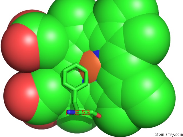
Mono view
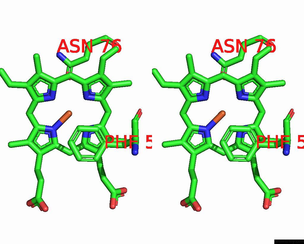
Stereo pair view
Mono view
Stereo pair view
A full contact list of Iron with other atoms in the Fe binding
site number 1 of Crystal Structure of the Wtblc-Split Protein within 5.0Å range:
|
Reference:
N.G.Bozhanova,
M.W.Calcutt,
W.N.Beavers,
B.P.Brown,
E.P.Skaar,
J.Meiler.
Lipocalin Blc Is A Potential Heme-Binding Protein. Febs Lett. 2020.
ISSN: ISSN 0014-5793
PubMed: 33210733
DOI: 10.1002/1873-3468.14001
Page generated: Wed Aug 7 13:34:02 2024
ISSN: ISSN 0014-5793
PubMed: 33210733
DOI: 10.1002/1873-3468.14001
Last articles
Zn in 9JYWZn in 9IR4
Zn in 9IR3
Zn in 9GMX
Zn in 9GMW
Zn in 9JEJ
Zn in 9ERF
Zn in 9ERE
Zn in 9EGV
Zn in 9EGW