Iron »
PDB 6vk8-6wnb »
6vw8 »
Iron in PDB 6vw8: Formate Dehydrogenase Fdsabg Subcomplex Fdsbg From C. Necator
Enzymatic activity of Formate Dehydrogenase Fdsabg Subcomplex Fdsbg From C. Necator
All present enzymatic activity of Formate Dehydrogenase Fdsabg Subcomplex Fdsbg From C. Necator:
1.2.1.2;
1.2.1.2;
Protein crystallography data
The structure of Formate Dehydrogenase Fdsabg Subcomplex Fdsbg From C. Necator, PDB code: 6vw8
was solved by
T.Young,
with X-Ray Crystallography technique. A brief refinement statistics is given in the table below:
| Resolution Low / High (Å) | 48.53 / 2.30 |
| Space group | C 1 2 1 |
| Cell size a, b, c (Å), α, β, γ (°) | 132.656, 127.050, 99.840, 90.00, 123.50, 90.00 |
| R / Rfree (%) | 18.2 / 22.3 |
Other elements in 6vw8:
The structure of Formate Dehydrogenase Fdsabg Subcomplex Fdsbg From C. Necator also contains other interesting chemical elements:
| Potassium | (K) | 2 atoms |
Iron Binding Sites:
Pages:
>>> Page 1 <<< Page 2, Binding sites: 11 - 12;Binding sites:
The binding sites of Iron atom in the Formate Dehydrogenase Fdsabg Subcomplex Fdsbg From C. Necator (pdb code 6vw8). This binding sites where shown within 5.0 Angstroms radius around Iron atom.In total 12 binding sites of Iron where determined in the Formate Dehydrogenase Fdsabg Subcomplex Fdsbg From C. Necator, PDB code: 6vw8:
Jump to Iron binding site number: 1; 2; 3; 4; 5; 6; 7; 8; 9; 10;
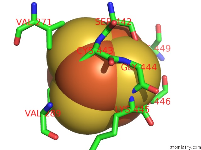
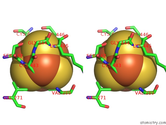
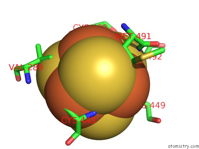
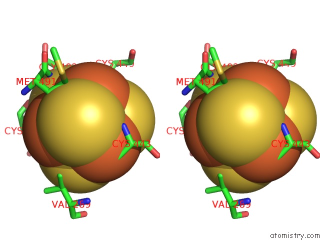
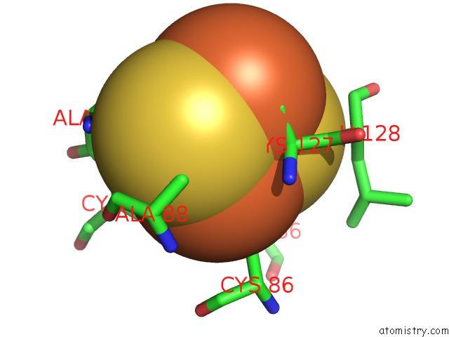
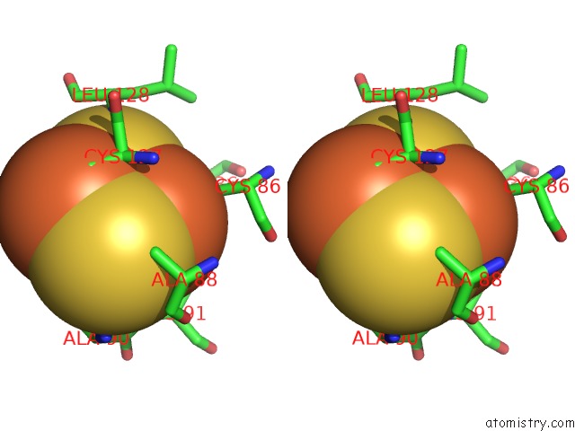
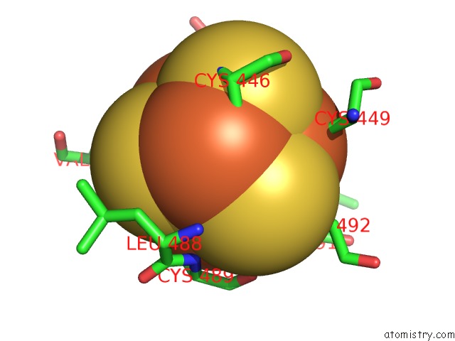
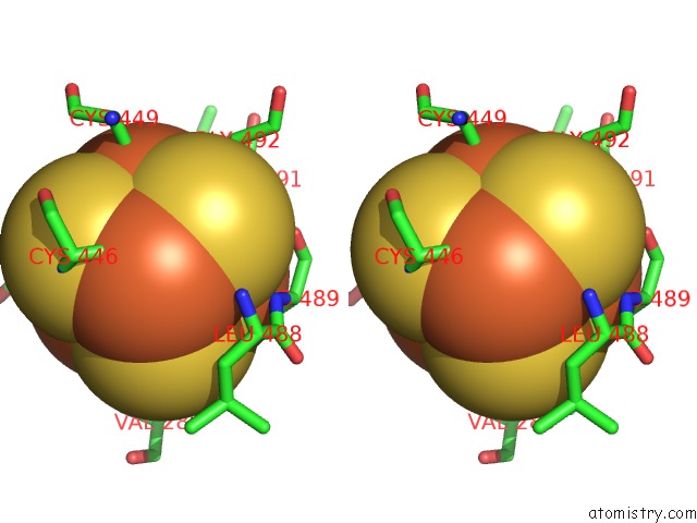
Iron binding site 1 out of 12 in 6vw8
Go back to
Iron binding site 1 out
of 12 in the Formate Dehydrogenase Fdsabg Subcomplex Fdsbg From C. Necator
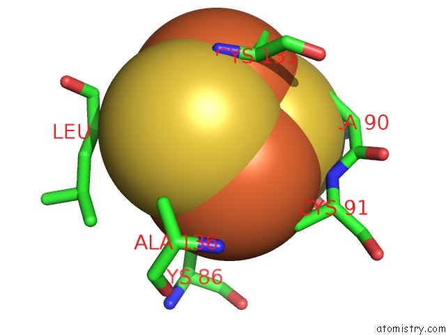
Mono view
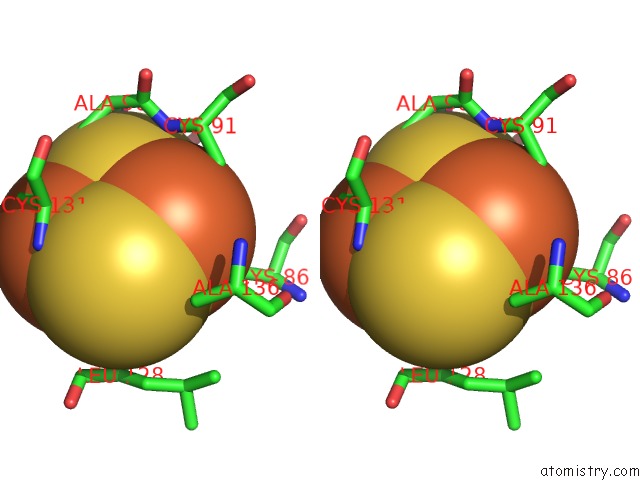
Stereo pair view
Mono view
Stereo pair view
A full contact list of Iron with other atoms in the Fe binding
site number 1 of Formate Dehydrogenase Fdsabg Subcomplex Fdsbg From C. Necator within 5.0Å range:
|
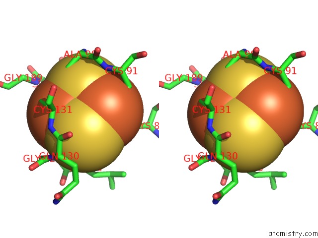
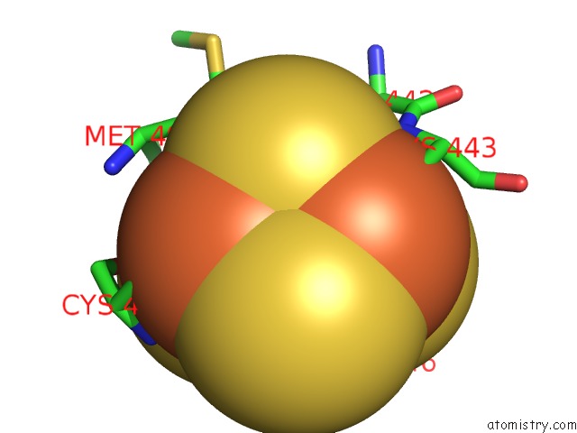
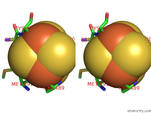
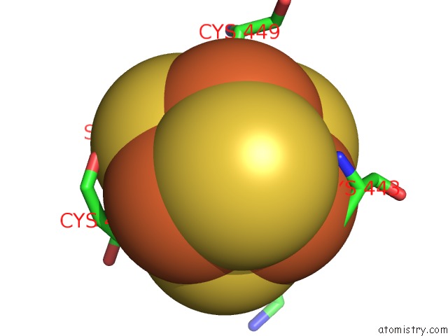
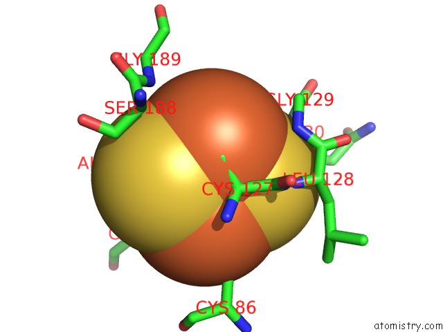
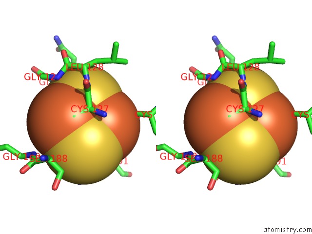
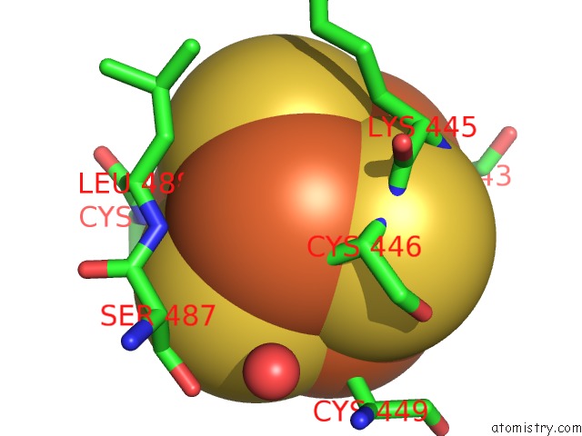
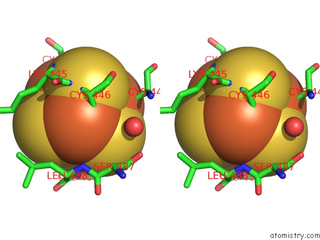
Iron binding site 2 out of 12 in 6vw8
Go back to
Iron binding site 2 out
of 12 in the Formate Dehydrogenase Fdsabg Subcomplex Fdsbg From C. Necator
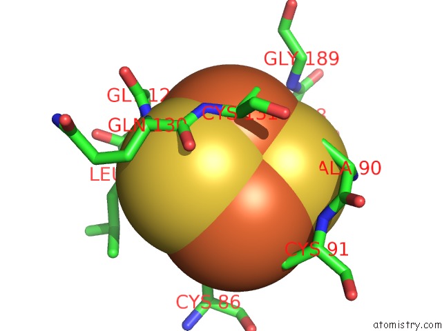
Mono view
Stereo pair view
Mono view
Stereo pair view
A full contact list of Iron with other atoms in the Fe binding
site number 2 of Formate Dehydrogenase Fdsabg Subcomplex Fdsbg From C. Necator within 5.0Å range:
|
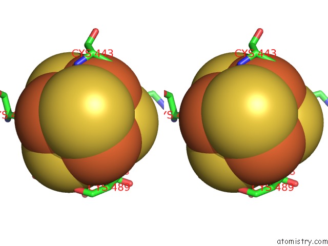
Iron binding site 3 out of 12 in 6vw8
Go back to
Iron binding site 3 out
of 12 in the Formate Dehydrogenase Fdsabg Subcomplex Fdsbg From C. Necator
Mono view
Stereo pair view
Mono view
Stereo pair view
A full contact list of Iron with other atoms in the Fe binding
site number 3 of Formate Dehydrogenase Fdsabg Subcomplex Fdsbg From C. Necator within 5.0Å range:
|
Iron binding site 4 out of 12 in 6vw8
Go back to
Iron binding site 4 out
of 12 in the Formate Dehydrogenase Fdsabg Subcomplex Fdsbg From C. Necator
Mono view
Stereo pair view
Mono view
Stereo pair view
A full contact list of Iron with other atoms in the Fe binding
site number 4 of Formate Dehydrogenase Fdsabg Subcomplex Fdsbg From C. Necator within 5.0Å range:
|
Iron binding site 5 out of 12 in 6vw8
Go back to
Iron binding site 5 out
of 12 in the Formate Dehydrogenase Fdsabg Subcomplex Fdsbg From C. Necator
Mono view
Stereo pair view
Mono view
Stereo pair view
A full contact list of Iron with other atoms in the Fe binding
site number 5 of Formate Dehydrogenase Fdsabg Subcomplex Fdsbg From C. Necator within 5.0Å range:
|
Iron binding site 6 out of 12 in 6vw8
Go back to
Iron binding site 6 out
of 12 in the Formate Dehydrogenase Fdsabg Subcomplex Fdsbg From C. Necator
Mono view
Stereo pair view
Mono view
Stereo pair view
A full contact list of Iron with other atoms in the Fe binding
site number 6 of Formate Dehydrogenase Fdsabg Subcomplex Fdsbg From C. Necator within 5.0Å range:
|
Iron binding site 7 out of 12 in 6vw8
Go back to
Iron binding site 7 out
of 12 in the Formate Dehydrogenase Fdsabg Subcomplex Fdsbg From C. Necator
Mono view
Stereo pair view
Mono view
Stereo pair view
A full contact list of Iron with other atoms in the Fe binding
site number 7 of Formate Dehydrogenase Fdsabg Subcomplex Fdsbg From C. Necator within 5.0Å range:
|
Iron binding site 8 out of 12 in 6vw8
Go back to
Iron binding site 8 out
of 12 in the Formate Dehydrogenase Fdsabg Subcomplex Fdsbg From C. Necator
Mono view
Stereo pair view
Mono view
Stereo pair view
A full contact list of Iron with other atoms in the Fe binding
site number 8 of Formate Dehydrogenase Fdsabg Subcomplex Fdsbg From C. Necator within 5.0Å range:
|
Iron binding site 9 out of 12 in 6vw8
Go back to
Iron binding site 9 out
of 12 in the Formate Dehydrogenase Fdsabg Subcomplex Fdsbg From C. Necator
Mono view
Stereo pair view
Mono view
Stereo pair view
A full contact list of Iron with other atoms in the Fe binding
site number 9 of Formate Dehydrogenase Fdsabg Subcomplex Fdsbg From C. Necator within 5.0Å range:
|
Iron binding site 10 out of 12 in 6vw8
Go back to
Iron binding site 10 out
of 12 in the Formate Dehydrogenase Fdsabg Subcomplex Fdsbg From C. Necator
Mono view
Stereo pair view
Mono view
Stereo pair view
A full contact list of Iron with other atoms in the Fe binding
site number 10 of Formate Dehydrogenase Fdsabg Subcomplex Fdsbg From C. Necator within 5.0Å range:
|
Reference:
T.Young,
D.Niks,
S.Hakopian,
T.K.Tam,
R.Hille,
G.Blaha.
Crystallographic and Kinetic Analyses of the Fdsbg Subcomplex of the Cytosolic Formate Dehydrogenase Fdsabg From Cupriavidus Necator Journal of Biological 2020CHEMISTRY.
Page generated: Wed Aug 7 13:37:28 2024
Last articles
Zn in 9MJ5Zn in 9HNW
Zn in 9G0L
Zn in 9FNE
Zn in 9DZN
Zn in 9E0I
Zn in 9D32
Zn in 9DAK
Zn in 8ZXC
Zn in 8ZUF