Iron »
PDB 6vk8-6wnb »
6wf2 »
Iron in PDB 6wf2: Crystal Structure of Mouse SCD1 with A Diiron Center
Enzymatic activity of Crystal Structure of Mouse SCD1 with A Diiron Center
All present enzymatic activity of Crystal Structure of Mouse SCD1 with A Diiron Center:
1.14.19.1;
1.14.19.1;
Protein crystallography data
The structure of Crystal Structure of Mouse SCD1 with A Diiron Center, PDB code: 6wf2
was solved by
J.Shen,
M.Zhou,
with X-Ray Crystallography technique. A brief refinement statistics is given in the table below:
| Resolution Low / High (Å) | 88.63 / 3.51 |
| Space group | P 21 21 21 |
| Cell size a, b, c (Å), α, β, γ (°) | 76.647, 113.981, 140.971, 90.00, 90.00, 90.00 |
| R / Rfree (%) | 21.9 / 27.7 |
Iron Binding Sites:
The binding sites of Iron atom in the Crystal Structure of Mouse SCD1 with A Diiron Center
(pdb code 6wf2). This binding sites where shown within
5.0 Angstroms radius around Iron atom.
In total 4 binding sites of Iron where determined in the Crystal Structure of Mouse SCD1 with A Diiron Center, PDB code: 6wf2:
Jump to Iron binding site number: 1; 2; 3; 4;
In total 4 binding sites of Iron where determined in the Crystal Structure of Mouse SCD1 with A Diiron Center, PDB code: 6wf2:
Jump to Iron binding site number: 1; 2; 3; 4;
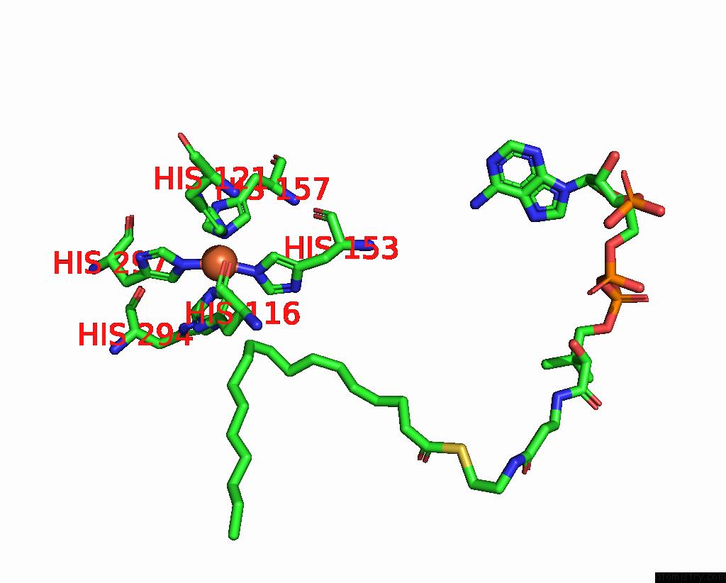
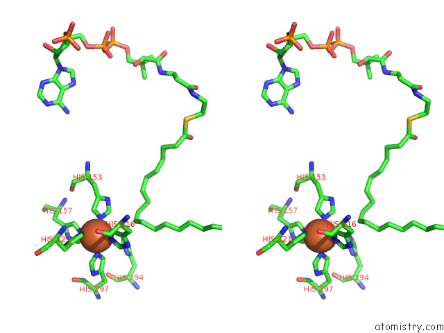
Iron binding site 1 out of 4 in 6wf2
Go back to
Iron binding site 1 out
of 4 in the Crystal Structure of Mouse SCD1 with A Diiron Center
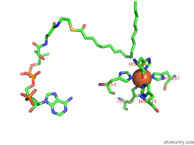
Mono view
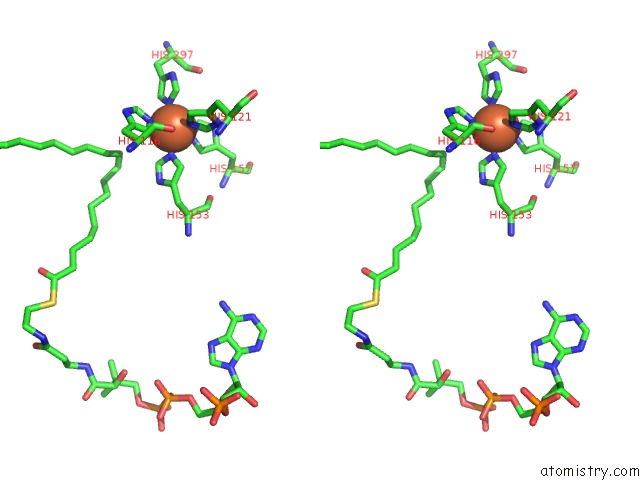
Stereo pair view
Mono view
Stereo pair view
A full contact list of Iron with other atoms in the Fe binding
site number 1 of Crystal Structure of Mouse SCD1 with A Diiron Center within 5.0Å range:
|
Iron binding site 2 out of 4 in 6wf2
Go back to
Iron binding site 2 out
of 4 in the Crystal Structure of Mouse SCD1 with A Diiron Center
Mono view
Stereo pair view
Mono view
Stereo pair view
A full contact list of Iron with other atoms in the Fe binding
site number 2 of Crystal Structure of Mouse SCD1 with A Diiron Center within 5.0Å range:
|
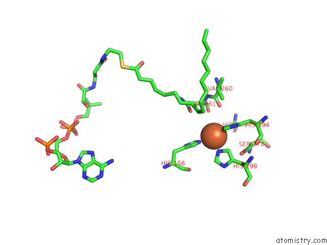
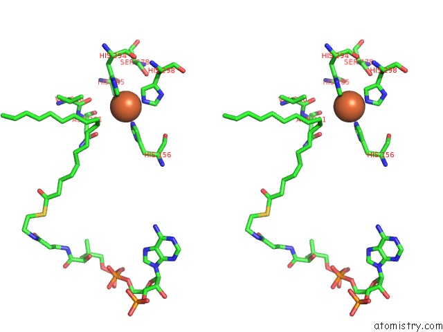
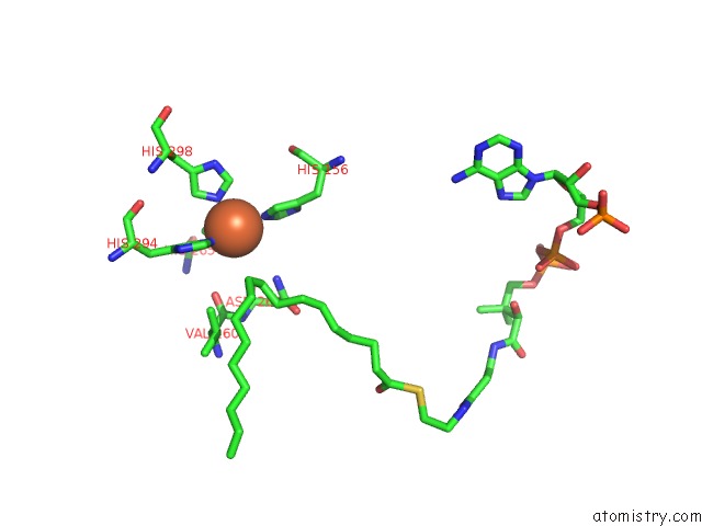
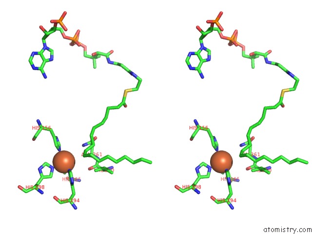
Iron binding site 3 out of 4 in 6wf2
Go back to
Iron binding site 3 out
of 4 in the Crystal Structure of Mouse SCD1 with A Diiron Center
Mono view
Stereo pair view
Mono view
Stereo pair view
A full contact list of Iron with other atoms in the Fe binding
site number 3 of Crystal Structure of Mouse SCD1 with A Diiron Center within 5.0Å range:
|
Iron binding site 4 out of 4 in 6wf2
Go back to
Iron binding site 4 out
of 4 in the Crystal Structure of Mouse SCD1 with A Diiron Center
Mono view
Stereo pair view
Mono view
Stereo pair view
A full contact list of Iron with other atoms in the Fe binding
site number 4 of Crystal Structure of Mouse SCD1 with A Diiron Center within 5.0Å range:
|
Reference:
J.Shen,
G.Wu,
A.L.Tsai,
M.Zhou.
Structure and Mechanism of A Unique Diiron Center in Mammalian Stearoyl-Coa Desaturase. J.Mol.Biol. 2020.
ISSN: ESSN 1089-8638
PubMed: 32470559
DOI: 10.1016/J.JMB.2020.05.017
Page generated: Wed Aug 7 13:58:58 2024
ISSN: ESSN 1089-8638
PubMed: 32470559
DOI: 10.1016/J.JMB.2020.05.017
Last articles
Cl in 2Z3JCl in 2Z2W
Cl in 2Z0R
Cl in 2Z2N
Cl in 2Z19
Cl in 2Z2K
Cl in 2Z0L
Cl in 2Z2D
Cl in 2Z18
Cl in 2Z12