Iron »
PDB 6wnc-6xa3 »
6x11 »
Iron in PDB 6x11: Observing A Ring-Cleaving Dioxygenase in Action Through A Crystalline Lens - An Enol Tautomer of Acms Monodentately Bound Structure
Enzymatic activity of Observing A Ring-Cleaving Dioxygenase in Action Through A Crystalline Lens - An Enol Tautomer of Acms Monodentately Bound Structure
All present enzymatic activity of Observing A Ring-Cleaving Dioxygenase in Action Through A Crystalline Lens - An Enol Tautomer of Acms Monodentately Bound Structure:
1.13.11.6;
1.13.11.6;
Protein crystallography data
The structure of Observing A Ring-Cleaving Dioxygenase in Action Through A Crystalline Lens - An Enol Tautomer of Acms Monodentately Bound Structure, PDB code: 6x11
was solved by
Y.Wang,
F.Liu,
Y.Yang,
A.Liu,
with X-Ray Crystallography technique. A brief refinement statistics is given in the table below:
| Resolution Low / High (Å) | 38.19 / 2.10 |
| Space group | P 65 2 2 |
| Cell size a, b, c (Å), α, β, γ (°) | 58.626, 58.626, 231.814, 90.00, 90.00, 120.00 |
| R / Rfree (%) | 21.9 / 27.9 |
Iron Binding Sites:
The binding sites of Iron atom in the Observing A Ring-Cleaving Dioxygenase in Action Through A Crystalline Lens - An Enol Tautomer of Acms Monodentately Bound Structure
(pdb code 6x11). This binding sites where shown within
5.0 Angstroms radius around Iron atom.
In total 2 binding sites of Iron where determined in the Observing A Ring-Cleaving Dioxygenase in Action Through A Crystalline Lens - An Enol Tautomer of Acms Monodentately Bound Structure, PDB code: 6x11:
Jump to Iron binding site number: 1; 2;
In total 2 binding sites of Iron where determined in the Observing A Ring-Cleaving Dioxygenase in Action Through A Crystalline Lens - An Enol Tautomer of Acms Monodentately Bound Structure, PDB code: 6x11:
Jump to Iron binding site number: 1; 2;
Iron binding site 1 out of 2 in 6x11
Go back to
Iron binding site 1 out
of 2 in the Observing A Ring-Cleaving Dioxygenase in Action Through A Crystalline Lens - An Enol Tautomer of Acms Monodentately Bound Structure
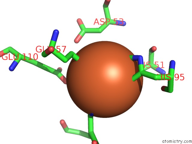
Mono view
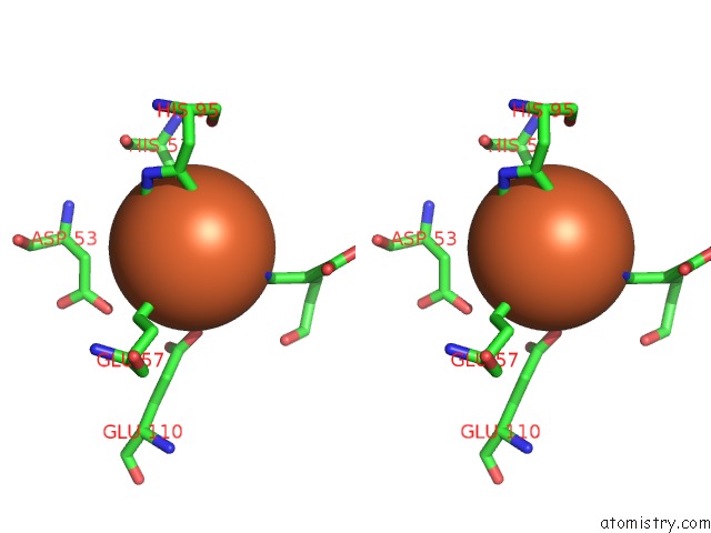
Stereo pair view
Mono view
Stereo pair view
A full contact list of Iron with other atoms in the Fe binding
site number 1 of Observing A Ring-Cleaving Dioxygenase in Action Through A Crystalline Lens - An Enol Tautomer of Acms Monodentately Bound Structure within 5.0Å range:
|
Iron binding site 2 out of 2 in 6x11
Go back to
Iron binding site 2 out
of 2 in the Observing A Ring-Cleaving Dioxygenase in Action Through A Crystalline Lens - An Enol Tautomer of Acms Monodentately Bound Structure
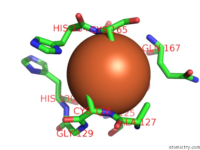
Mono view
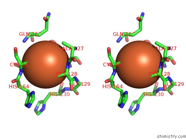
Stereo pair view
Mono view
Stereo pair view
A full contact list of Iron with other atoms in the Fe binding
site number 2 of Observing A Ring-Cleaving Dioxygenase in Action Through A Crystalline Lens - An Enol Tautomer of Acms Monodentately Bound Structure within 5.0Å range:
|
Reference:
Y.Wang,
F.Liu,
Y.Yang,
A.Liu.
Probing Extradiol Dioxygenase Mechanism in Nad+ Biosynthesis By Viewing Reaction Cycle Intermediates Proc.Natl.Acad.Sci.Usa 2020.
ISSN: ESSN 1091-6490
DOI: 10.1073/PNAS.2005327117
Page generated: Wed Aug 7 14:37:26 2024
ISSN: ESSN 1091-6490
DOI: 10.1073/PNAS.2005327117
Last articles
Zn in 9J0NZn in 9J0O
Zn in 9J0P
Zn in 9FJX
Zn in 9EKB
Zn in 9C0F
Zn in 9CAH
Zn in 9CH0
Zn in 9CH3
Zn in 9CH1